surgeonring

The anatomy journal

The body is a living story.

Surgeonring explains anatomy, physiology, biology, and histology as one connected subject — every organ traced from its position in the body down to the cells that make it work.
12 organ guidesCited from standard textsWritten to be re-read

The living index / 12 studies

Choose an organ.
Follow the system.

Every guide moves from visible structure to microscopic tissue, then answers the question that matters most: how does it actually work?

01 / 12
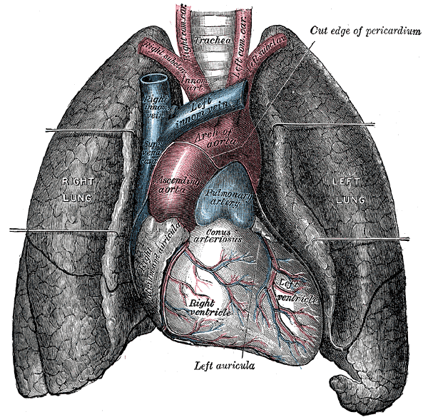
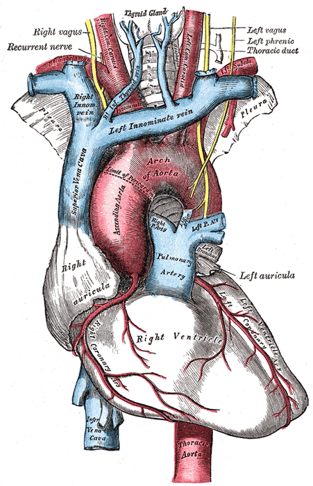
The heart and great vessels in situ, seen from the front with the lungs retracted.

Cardiovascular system / 7 min read

Heart

A complete guide to the human heart: chambers, valves and coronary vessels, the cardiac cycle, excitation-contraction coupling, cardiomyocyte biology, and the histology of all three cardiac layers.

Open study
02 / 12
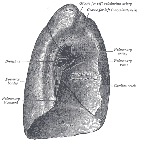
Mediastinal surface of the left lung, showing the hilum and cardiac impression.

Respiratory system / 6 min read

Lungs

How the lungs are built and how they work: the bronchial tree, pleura and respiratory mechanics, gas exchange and oxygen transport, alveolar cell biology, and the histology of the respiratory membrane.

Open study
03 / 12
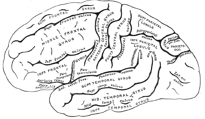
Lateral surface of the cerebral hemisphere, with the principal gyri and sulci named.

Nervous system / 6 min read

Brain

A structured guide to the human brain: lobes and deep structures, the meninges and cerebrospinal fluid, synaptic transmission, neuronal and glial biology, and the histology of grey and white matter.

Open study
04 / 12
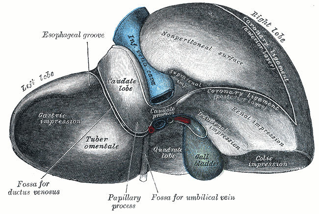
Visceral surface of the liver, showing the lobes, gallbladder and porta hepatis.

Digestive system / 7 min read

Liver

The liver explained in depth: lobes and segments, dual blood supply, metabolic and detoxification physiology, hepatocyte biology, the classic lobule and acinus, and the pathology of cirrhosis.

Open study
05 / 12
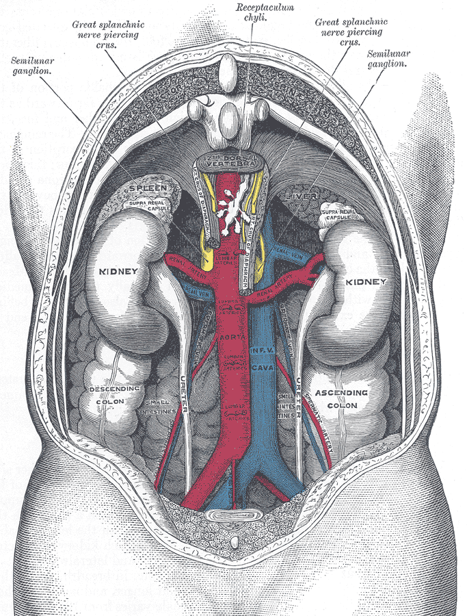
The kidneys and great vessels in situ on the posterior abdominal wall.

Urinary system / 7 min read

Kidneys

Follow blood through the nephron: renal anatomy, glomerular filtration, tubular transport, the countercurrent multiplier, hormonal control of volume and pressure, and renal histology segment by segment.

Open study
06 / 12
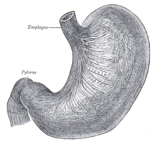
The stomach seen from the front, from the cardia through the body to the pylorus.

Digestive system / 7 min read

Stomach

How the stomach works: regions and wall layers, acid secretion by parietal cells, the three phases of gastric secretion, gastric gland biology, and the mucosal barrier that stops the organ digesting itself.

Open study
07 / 12
The caecum, ileum and ascending colon with their arterial supply.

Digestive system / 7 min read

Intestines

The small and large intestine explained: regional anatomy, the surface amplification that makes absorption possible, motility patterns, enterocyte biology, the microbiome, and mucosal histology.

Open study
08 / 12
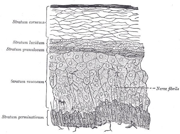
Vertical section through the skin, showing epidermis, dermis and the appendages.

Integumentary system / 7 min read

Skin

The skin as a working organ: epidermal layers and keratinocyte maturation, dermal connective tissue and appendages, barrier function, thermoregulation, sensation, wound healing, and skin histology.

Open study
09 / 12
The pancreas and duodenum, showing the main and accessory pancreatic ducts.

Digestive and endocrine systems / 7 min read

Pancreas

One organ, two systems: pancreatic anatomy and ducts, enzyme and bicarbonate secretion, insulin and glucagon control of blood glucose, acinar and islet cell biology, and pancreatic histology.

Open study
10 / 12
The spleen, visceral surface, showing the hilum and splenic vessels.

Lymphatic and immune systems / 7 min read

Spleen

The spleen as a blood filter: position and relations, open and closed circulation, red pulp quality control of erythrocytes, white pulp immune surveillance, splenic histology, and the risks of splenectomy.

Open study
11 / 12
The thyroid gland seen from behind, with the parathyroid glands and inferior thyroid artery.

Endocrine system / 7 min read

Thyroid

How the thyroid sets metabolic pace: neck anatomy and surgical relations, iodine trapping and hormone synthesis, the hypothalamic-pituitary-thyroid axis, follicular cell biology, and thyroid histology.

Open study
12 / 12
The knee joint opened from the front, showing the femoral condyles and cruciate ligaments.

Skeletal system / 8 min read

Bone

Bone as living tissue: gross structure and classification, the mineral and collagen composite, calcium homeostasis, osteoblast and osteoclast biology, osteon histology, remodelling, and fracture repair.

Open study

A better way to look

From the whole organ to the living cell.

A body is not a collection of flashcards. Each Surgeonring study moves through four lenses in the same order, so that structure explains function instead of sitting beside it.

  • Anatomy

    Where the organ sits, what it is attached to, and which vessels and nerves reach it.

  • Physiology

    The mechanism: what the organ actually does, step by step, and what regulates it.

  • Biology

    The cells that do the work, and why each one is shaped for the job it holds.

  • Histology

    What the tissue looks like under magnification, and how to recognise it on a slide.