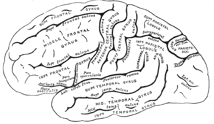
The brain weighs about 1.4 kilograms, roughly two percent of body mass, yet consumes close to twenty percent of the body's oxygen and glucose at rest. It has no meaningful fuel reserve, which is why consciousness is lost within seconds of interrupted blood flow. Its complexity is real, but its organising principle is simple: specialised cells communicate across connected regions, and behaviour emerges from the pattern of those connections rather than from any single location.
Anatomy: the structure
The cerebrum is divided into two hemispheres joined by the corpus callosum, and each hemisphere carries frontal, parietal, temporal, and occipital lobes plus the deeply buried insula. The frontal lobe holds the primary motor cortex and the executive and language production regions, the parietal lobe the primary somatosensory cortex, the temporal lobe auditory processing and the hippocampal memory system, and the occipital lobe the primary visual cortex. Folding into gyri and sulci roughly triples the cortical surface area that fits inside the skull.
Beneath the cortex sit the basal ganglia, which shape the initiation and scaling of movement, the thalamus, which relays almost all sensory traffic to the cortex, and the hypothalamus, which governs temperature, hunger, thirst, circadian timing, and endocrine output through the pituitary. The cerebellum, behind the brainstem, coordinates timing, balance, and motor learning. The brainstem, comprising midbrain, pons, and medulla, carries the cranial nerve nuclei and the autonomic centres that control breathing and cardiovascular tone.
Three protective layers surround the brain. The tough dura mater lines the skull and folds inward as the falx cerebri and tentorium cerebelli, the delicate arachnoid mater spans the surface, and the pia mater follows every contour of the cortex. Cerebrospinal fluid produced by the choroid plexus circulates through the ventricles and subarachnoid space, cushioning the brain and effectively reducing its weight. Blood arrives through the paired internal carotid and vertebral arteries, which anastomose in the circle of Willis, and the blood-brain barrier formed by tight junctions between capillary endothelial cells tightly controls what crosses into neural tissue.
Physiology: how it works
Neurons maintain a resting membrane potential near minus 70 millivolts using the sodium-potassium ATPase and selective ion permeability. When summed excitatory input at the axon hillock reaches threshold, voltage-gated sodium channels open and an all-or-nothing action potential propagates along the axon. Myelination allows saltatory conduction between nodes of Ranvier, raising conduction velocity from around one metre per second to over one hundred.
At the synapse, arriving depolarisation opens voltage-gated calcium channels, vesicles fuse with the presynaptic membrane, and neurotransmitter diffuses across the cleft to bind postsynaptic receptors. Glutamate is the principal excitatory transmitter and GABA the principal inhibitory one, while dopamine, serotonin, acetylcholine, and noradrenaline act as modulators that bias whole networks. A single cortical neuron may receive thousands of such inputs and continuously integrate them in space and time.
Function is distributed rather than localised. Sensory information is processed hierarchically, from primary cortex through association areas, and motor output is planned in premotor regions before the primary motor cortex issues commands. Synaptic strength changes with use through long-term potentiation and depression, which is the cellular basis of learning. Meanwhile the autonomic hierarchy from hypothalamus to brainstem regulates heart rate, blood pressure, temperature, appetite, and the sleep-wake cycle without conscious involvement.
Biology: the living cells
The adult brain contains roughly 86 billion neurons and a comparable number of glial cells. A typical neuron has dendrites that receive input, a soma containing the nucleus and abundant rough endoplasmic reticulum visible as Nissl substance, and a single axon that carries output. Glia are not passive packing: astrocytes buffer extracellular potassium, recycle glutamate, supply metabolic substrate, and help induce the blood-brain barrier; oligodendrocytes myelinate multiple central axons each, while Schwann cells do the same job one internode at a time in the peripheral nervous system; microglia are resident immune cells derived from the yolk sac that survey tissue and prune synapses; and ependymal cells line the ventricles and move cerebrospinal fluid with their cilia. Because mature neurons are largely postmitotic, most of the brain's adaptability comes from changing connections rather than from making new cells.
Histology: under the microscope
Nervous tissue separates into grey matter, rich in neuronal cell bodies, dendrites, synapses, and unmyelinated processes, and white matter, dominated by myelinated axon tracts whose lipid content gives the pale colour. The cerebral cortex is classically organised into six layers that differ in cell type and connectivity, with layer IV enlarged in sensory cortex and layer V enlarged in motor cortex where the large pyramidal Betz cells sit. The cerebellar cortex has its own three-layer arrangement of molecular, Purkinje, and granular layers, and the enormous dendritic tree of the Purkinje cell is one of the most recognisable images in histology. Silver and immunohistochemical stains reveal what routine staining hides: the dense, branching neuropil in which almost all synaptic contact occurs.
Clinical correlations
Ischaemic stroke follows occlusion of a cerebral artery, and because the brain has no substrate reserve, the core infarct dies within minutes while the surrounding penumbra remains salvageable for a few hours. The clinical deficit maps predictably onto the vessel involved: middle cerebral artery occlusion typically produces contralateral face and arm weakness with aphasia when the dominant hemisphere is affected, while anterior cerebral artery occlusion affects the leg disproportionately.
Neurodegenerative disease is defined by which cell population fails. Alzheimer disease involves amyloid plaques and tau neurofibrillary tangles with early hippocampal and entorhinal involvement, producing memory loss first. Parkinson disease follows loss of dopaminergic neurons in the substantia nigra pars compacta, disrupting basal ganglia output and producing bradykinesia, rigidity, and tremor. Multiple sclerosis is a demyelinating disease in which immune attack on oligodendrocyte myelin slows or blocks conduction, generating deficits separated in time and location.
Because the skull is a rigid container, the Monro-Kellie doctrine means that any added volume from haemorrhage, tumour, or oedema must displace brain, blood, or cerebrospinal fluid, and once compensation is exhausted intracranial pressure rises steeply and herniation threatens the brainstem. Epilepsy, by contrast, is a disorder of excitability rather than volume, arising when the balance between glutamatergic excitation and GABAergic inhibition tips toward synchronous discharge.
Common questions about the brain
What is the difference between grey matter and white matter?
Grey matter contains neuronal cell bodies, dendrites, and synapses and is where most information processing occurs. White matter consists mainly of myelinated axons that connect regions to one another, and the pale colour comes from the lipid-rich myelin surrounding those axons.
How do neurons communicate with each other?
An action potential travelling down an axon opens calcium channels at the terminal, causing vesicles to release neurotransmitter into the synaptic cleft. The transmitter binds receptors on the next neuron, making it either more or less likely to fire, and the receiving neuron sums thousands of such inputs before deciding whether to generate its own action potential.
What does the blood-brain barrier actually do?
Tight junctions between brain capillary endothelial cells, supported by astrocyte end-feet and pericytes, prevent free passage of most substances from blood into neural tissue. This keeps the extracellular environment chemically stable and excludes many toxins and pathogens, but it also blocks a large fraction of otherwise useful drugs.
Can the adult brain make new neurons?
Most adult neurons are postmitotic and are not replaced. Limited neurogenesis has been described in the hippocampal dentate gyrus, but its extent in adult humans remains debated. Functional recovery after injury depends far more on synaptic reorganisation and changing connection strength than on producing new cells.
Selected references
- Kandel ER, Koester JD, Mack SH, Siegelbaum SA. Principles of Neural Science. 6th ed. McGraw Hill; 2021.
- Standring S, ed. Gray's Anatomy: The Anatomical Basis of Clinical Practice. 42nd ed. Elsevier; 2021.
- Purves D, et al. Neuroscience. 6th ed. Oxford University Press; 2018.
- Azevedo FAC, et al. Equal numbers of neuronal and nonneuronal cells make the human brain an isometrically scaled-up primate brain. Journal of Comparative Neurology. 2009;513(5):532-541.
- Ross MH, Pawlina W. Histology: A Text and Atlas. 8th ed. Wolters Kluwer; 2020.
- Blumenfeld H. Neuroanatomy through Clinical Cases. 3rd ed. Sinauer; 2021.
Medical disclaimer. This article is written for education and general understanding. It is not medical advice and cannot replace assessment by a qualified healthcare professional. If you have symptoms or health concerns, speak with a clinician. Read the full disclaimer.