An adult carries about five litres of blood, and if you spin a sample of it in a centrifuge it separates cleanly into the answer to the question. Roughly 55 per cent is plasma, a straw-coloured liquid that is mostly water. Roughly 45 per cent is red cells, packed at the bottom. Between them sits a thin pale band, less than one per cent of the total, called the buffy coat, holding all the white cells and platelets. That last sliver does the immune defence and the clotting for the entire body. Blood is classified as a connective tissue, which surprises people, but it fits the definition exactly: scattered cells suspended in a matrix they did not make and do not touch.
Where it is, and what it looks like
Plasma is about 92 per cent water and 7 per cent protein, with the remaining one per cent made up of electrolytes, nutrients, hormones, dissolved gases and waste. The proteins matter more than their small share suggests. Albumin, made in the liver, is more than half of the total and provides the colloid osmotic pressure that holds water inside the vessels — which is why liver failure and severe malnutrition both cause oedema. Globulins include the antibodies and the transport proteins for iron, copper and hormones. Fibrinogen is the soluble precursor that becomes the fibrin mesh of a clot; serum is simply plasma with the fibrinogen used up.
Red cells, or erythrocytes, are the most numerous cells in the body at roughly 25 trillion in an adult, around 4.5 to 6 million per microlitre of blood. Each is a biconcave disc about 7 to 8 micrometres across, with no nucleus and no mitochondria, packed with roughly 270 million molecules of haemoglobin. Losing the nucleus frees space for haemoglobin and gives the cell the flexibility to fold through capillaries narrower than itself; losing the mitochondria means the cell cannot consume the oxygen it is carrying, relying on anaerobic glycolysis instead. The cost of having no nucleus is that the cell cannot repair itself, so it wears out after about 120 days and is removed by macrophages in the spleen and liver.
White cells, or leukocytes, number only 4,000 to 11,000 per microlitre and divide into two families. The granulocytes — neutrophils, eosinophils and basophils — have lobed nuclei and cytoplasmic granules. Neutrophils are the most abundant white cell at 40 to 75 per cent and are the first responders to bacterial infection. Eosinophils handle parasites and drive allergic inflammation; basophils release histamine and heparin. The agranulocytes are lymphocytes, at 20 to 45 per cent, which run the adaptive immune response as B, T and natural killer cells, and monocytes, which leave the blood to become tissue macrophages. Platelets are not cells at all but membrane-bound fragments shed from megakaryocytes, 150,000 to 400,000 per microlitre, and they live about eight to ten days.
What it does
The first function is transport, and almost all of it is oxygen. Haemoglobin carries roughly 98.5 per cent of the oxygen in blood, with the rest dissolved in plasma, and its four subunits bind oxygen cooperatively — each one that binds makes the next bind more easily, which produces the sigmoid dissociation curve and is what allows blood to load oxygen almost fully in the lungs and unload it readily in the tissues. That curve shifts right, releasing more oxygen, wherever tissue is hot, acidic, or high in carbon dioxide, meaning exercising muscle automatically extracts more. Carbon dioxide travels back mostly as bicarbonate generated inside the red cell by carbonic anhydrase, which makes red cells central to acid-base balance as well as to oxygen delivery.
The second function is defence. Neutrophils reach a site of infection within minutes, squeezing between endothelial cells and following chemical gradients, then engulf bacteria and kill them with reactive oxygen species and enzymes, dying in the process — pus is largely spent neutrophils. Monocytes arrive later and become macrophages, which clear debris and present antigen to lymphocytes. Lymphocytes provide the specific response: B cells produce antibodies, T cells kill infected cells and coordinate the whole reaction, and both leave behind memory. Because these populations respond differently, the differential white cell count is genuinely informative — a neutrophil rise suggests bacterial infection, a lymphocyte rise suggests viral, and an eosinophil rise suggests parasites or allergy.
The third function is haemostasis, and it runs in three overlapping stages. Injured vessels constrict immediately. Platelets then adhere to exposed collagen through von Willebrand factor, activate, change shape, release their granules and recruit more platelets into a plug — primary haemostasis, sufficient for small injuries. In parallel the coagulation cascade converts prothrombin to thrombin, and thrombin converts soluble fibrinogen into insoluble fibrin strands that mesh through the platelet plug and trap red cells, producing a stable clot. Blood also stabilises the body's temperature, pH and fluid balance, distributing heat from the core to the skin and buffering acid produced by metabolism.
The cells that build it
Every blood cell in the body descends from a single population of haematopoietic stem cells in the red bone marrow — in an adult, mainly in the pelvis, sternum, vertebrae, ribs and the ends of the femur and humerus. Those stem cells are both self-renewing and multipotent: one daughter stays a stem cell, the other commits to a lineage. The myeloid line gives rise to red cells, platelets via megakaryocytes, and all the granulocytes and monocytes; the lymphoid line gives rise to B, T and natural killer cells. Which path a cell takes is directed by growth factors, and the two best known are used as drugs: erythropoietin, made by the kidney in response to low oxygen, drives red cell production, and granulocyte colony-stimulating factor drives neutrophil production. Red cell development is a progressive loss of everything not needed for carrying oxygen — the cell accumulates haemoglobin, then extrudes its nucleus, leaving a reticulocyte that still contains ribosomal RNA and matures in the circulation over a day or two. The reticulocyte count is therefore a direct readout of how hard the marrow is working. Roughly two million red cells are produced and destroyed every second in a healthy adult, and the iron released when old cells are broken down in the spleen is recycled almost completely, which is why dietary iron requirements are far smaller than the amount of iron actually used each day.
How to recognise it on a slide
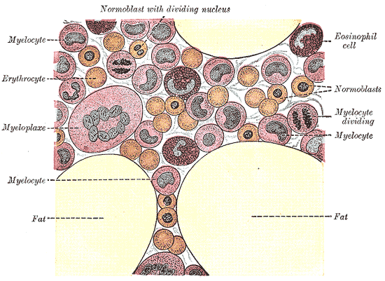
A blood film is one of the few preparations where the cells are spread in a single layer rather than sectioned, and the stain used is a Romanowsky stain such as Wright's or Giemsa rather than routine haematoxylin and eosin. Red cells dominate the field as pink discs with a pale centre, because the biconcave shape is thinner in the middle — that central pallor should be less than a third of the diameter, and a larger one suggests iron deficiency. Platelets appear as tiny purple fragments, often clumped, easily mistaken for debris. The white cells are identified by their nuclei. A neutrophil has a nucleus of three to five lobes joined by thin strands and pale granules; an eosinophil has a bilobed nucleus and large bright orange-red granules that are impossible to miss; a basophil is rare and so densely packed with dark purple granules that the nucleus is obscured. A lymphocyte is small, with a round dark nucleus filling almost the entire cell and only a thin rim of blue cytoplasm. A monocyte is the largest white cell, with an indented kidney-shaped or horseshoe nucleus and abundant greyish cytoplasm. Bone marrow, by contrast, shows the whole developmental sequence at once, with cells at every stage packed between fat spaces and sinusoids.
When it goes wrong
Anaemia is a fall in haemoglobin rather than in cell number, and its causes are best sorted by red cell size. Microcytic anaemia, with small pale cells, is usually iron deficiency, and in an adult the important question is where the iron is being lost, since chronic gastrointestinal bleeding may be the first sign of a colonic cancer. Macrocytic anaemia, with large cells, points to vitamin B12 or folate deficiency, in which DNA synthesis fails while cytoplasm continues to accumulate. Normocytic anaemia accompanies chronic disease, kidney failure through loss of erythropoietin, and acute blood loss. Sickle cell disease and thalassaemia are inherited faults in the haemoglobin molecule itself.
Disorders of white cells run in both directions. Leukaemia is uncontrolled proliferation of a white cell line in the marrow, and its danger comes as much from what it crowds out as from the abnormal cells themselves: the patient becomes anaemic, thrombocytopenic and, despite a high white cell count, functionally immunosuppressed. Neutropenia, whether from chemotherapy or marrow failure, removes the first line of antibacterial defence, and a fever in a neutropenic patient is a medical emergency treated with antibiotics before any organism is identified. Lymphoma is the same class of problem arising in lymphoid tissue rather than in the marrow.
Platelet and clotting disorders also fail in both directions. Too few platelets, or platelets that do not work, produces bleeding into the skin and mucous membranes — petechiae, bruising, nosebleeds and heavy periods. Haemophilia, a deficiency of clotting factor VIII or IX, produces the opposite pattern: bleeding into joints and muscles, because primary haemostasis works but the clot is never stabilised. At the other end, excessive clotting causes deep vein thrombosis and pulmonary embolism, and in disseminated intravascular coagulation the two extremes occur together, as widespread microvascular clotting consumes platelets and clotting factors until the patient bleeds.
Common questions about blood
What is blood made of?
Blood has four components. Plasma, about 55 per cent of the volume, is water carrying albumin, globulins, fibrinogen, electrolytes, nutrients, hormones and waste. Red cells, about 45 per cent, carry oxygen using haemoglobin. White cells provide immune defence and come in five types: neutrophils, lymphocytes, monocytes, eosinophils and basophils. Platelets are cell fragments that start the clotting process. White cells and platelets together make up less than one per cent of the volume.
Why is blood classed as a connective tissue?
Because it fits the definition: cells scattered in an extracellular matrix that they do not touch and did not make. In blood the matrix is liquid plasma rather than a gel or a mineralised solid, and the fibres exist only in potential, appearing as fibrin when clotting is triggered. It is grouped with cartilage and bone as a specialised connective tissue for exactly this reason.
What are the different types of white blood cells and what do they do?
Neutrophils, the most numerous, are the first responders to bacterial infection and die doing it. Lymphocytes run the specific immune response as B cells producing antibodies, T cells killing infected cells and directing the response, and natural killer cells. Monocytes leave the blood and become tissue macrophages. Eosinophils deal with parasites and drive allergic inflammation. Basophils release histamine and heparin. Which type is raised on a blood count is a genuine clue to what kind of illness is present.
Where are blood cells made?
In the red bone marrow, from haematopoietic stem cells. In an adult this means mainly the pelvis, sternum, vertebrae, ribs, and the ends of the femur and humerus; in a child, almost every bone. All blood cells descend from the same stem cell population, which splits into a myeloid line producing red cells, platelets, granulocytes and monocytes, and a lymphoid line producing B, T and natural killer cells. T lymphocytes complete their maturation in the thymus rather than the marrow.
Selected references
- Hoffbrand AV, Moss PAH. Hoffbrand's Essential Haematology. 8th ed. Wiley-Blackwell; 2019.
- Hall JE, Hall ME. Guyton and Hall Textbook of Medical Physiology. 14th ed. Elsevier; 2021.
- Ross MH, Pawlina W. Histology: A Text and Atlas. 8th ed. Wolters Kluwer; 2020.
- Sender R, Fuchs S, Milo R. Revised estimates for the number of human and bacteria cells in the body. PLoS Biology. 2016;14(8):e1002533.
- Orkin SH, Zon LI. Hematopoiesis: an evolving paradigm for stem cell biology. Cell. 2008;132(4):631-644.
- Kumar V, Abbas AK, Aster JC. Robbins and Cotran Pathologic Basis of Disease. 10th ed. Elsevier; 2021.
Medical disclaimer. This article is written for education and general understanding. It is not medical advice and cannot replace assessment by a qualified healthcare professional. If you have symptoms or health concerns, speak with a clinician. Read the full disclaimer.