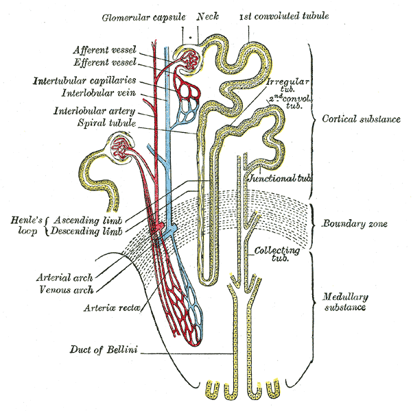
The kidney's strategy is strange when you first meet it. Rather than picking waste out of the blood, it throws almost everything away and then carefully takes back what it wants. Each of your roughly one million nephrons per kidney filters plasma indiscriminately, producing about 180 litres of filtrate a day, and then reclaims more than 99 per cent of it, leaving one to two litres of urine. It looks wasteful and it is not: filtering first and choosing afterwards means the body can regulate the composition of blood continuously by adjusting what it takes back, rather than by identifying every possible waste product in advance.
Where it is, and what it looks like
A nephron has two parts: a renal corpuscle that filters and a long tubule that processes. The corpuscle consists of the glomerulus, a tuft of capillaries, cupped inside Bowman's capsule. Blood enters the tuft through an afferent arteriole and leaves through an efferent arteriole — a capillary bed sitting between two arterioles rather than between an arteriole and a venule, which is what allows the kidney to control the pressure inside it from both ends. Beyond the corpuscle, the tubule runs as the proximal convoluted tubule, the loop of Henle with its descending and ascending limbs, the distal convoluted tubule, and finally the collecting duct, which several nephrons share.
The two ends of the nephron sit in different places, and this matters. The corpuscles and both convoluted tubules lie in the cortex; the loops of Henle and collecting ducts descend into the medulla. About 85 per cent of nephrons are cortical, with short loops that barely enter the medulla. The other 15 per cent are juxtamedullary, sitting deep in the cortex with long loops reaching far into the medulla, and they are the ones that generate the concentration gradient which makes concentrated urine possible. Their efferent arterioles form the vasa recta, long straight capillaries that follow the loops down and back.
Where the ascending limb returns to its own glomerulus it forms the juxtaglomerular apparatus, a small structure that does the kidney's self-regulation. The macula densa is a patch of closely packed tubular cells that senses sodium chloride concentration in the filtrate; the juxtaglomerular cells are modified smooth muscle in the wall of the afferent arteriole that store and release renin. Together they let each nephron adjust its own filtration rate and, through renin, influence blood pressure across the whole body.
What it does
Filtration is the first step and it is purely physical. Blood pressure in the glomerular capillaries, around 55 millimetres of mercury and much higher than in an ordinary capillary bed, pushes fluid across a three-layered barrier: fenestrated endothelium, a thick basement membrane, and the interdigitating foot processes of podocytes, whose narrow filtration slits are the final sieve. Opposing it are capsular hydrostatic pressure and the colloid osmotic pressure of the plasma proteins left behind, giving a net filtration pressure of about 10 millimetres of mercury. Water, ions, glucose, amino acids and urea cross freely; cells and plasma proteins do not, both because they are too large and because the basement membrane carries a fixed negative charge that repels albumin. The result is a glomerular filtration rate of roughly 125 millilitres per minute, or 180 litres a day.
Reabsorption is the second step, and most of it happens immediately. The proximal convoluted tubule reclaims about two-thirds of the filtered water and sodium, and essentially all of the glucose, amino acids and bicarbonate, using sodium gradients created by basolateral sodium-potassium pumps to drive secondary active transport at the apical brush border. Because glucose is reabsorbed by a finite number of transporters, exceeding their capacity — at a plasma glucose of roughly 10 to 11 millimoles per litre — causes glucose to appear in the urine, which is why uncontrolled diabetes produces sugary urine and, because that glucose drags water with it, thirst and high urine volumes.
The loop of Henle then builds the machinery for concentrating urine, by countercurrent multiplication. The descending limb is permeable to water but not to salt, so filtrate passing down it loses water into the increasingly salty medulla and becomes concentrated. The thick ascending limb is the reverse, impermeable to water but actively pumping sodium, potassium and chloride out through the NKCC2 transporter, so the filtrate leaving it is dilute while the surrounding medulla is made progressively saltier, reaching around 1,200 milliosmoles per kilogram at the tip. The vasa recta preserve that gradient by countercurrent exchange. Finally the distal tubule and collecting duct fine-tune everything under hormonal control: aldosterone increases sodium reabsorption and potassium secretion, antidiuretic hormone inserts aquaporin-2 channels that let water leave the collecting duct into the salty medulla, and parathyroid hormone adjusts calcium. The tubule also secretes — actively adding hydrogen ions, potassium, creatinine and many drugs into the filtrate — which is a second route for clearing substances the glomerulus did not filter efficiently.
The cells that build it
Each segment of the tubule is lined by a cell type built for its specific job, and you can predict the histology from the physiology. Proximal tubule cells are the workhorses: tall cuboidal, with a dense apical brush border of microvilli that multiplies the absorptive surface many times over, enormous numbers of mitochondria packed into deep basolateral membrane infoldings to power the sodium pumps, and extensive endocytic machinery to retrieve the small amount of protein that escapes filtration. That metabolic demand, combined with sitting at the end of a long low-oxygen blood supply, is exactly why proximal tubule cells are the first to die in shock and the classic site of acute tubular necrosis. The thin descending limb is simple squamous with few mitochondria, because it does nothing but let water leave passively. The thick ascending limb returns to cuboidal cells rich in mitochondria to run the NKCC2 pump, the transporter that loop diuretics block. The collecting duct holds two distinct populations: principal cells, which carry the aldosterone and antidiuretic hormone receptors and handle sodium, potassium and water, and intercalated cells, which secrete hydrogen ions or bicarbonate and are the final arbiter of blood pH. Podocytes deserve a note of their own: they are terminally differentiated, cannot divide, and once lost are not replaced, which is a central reason why glomerular damage tends to be permanent.
How to recognise it on a slide
A section of renal cortex is one of the most recognisable slides in histology. Renal corpuscles appear as round balls of capillaries surrounded by a clear space, Bowman's space, and a thin parietal layer of simple squamous epithelium; you can often identify the vascular pole, where the arterioles enter, and the urinary pole, where the proximal tubule begins. Scattered between them are two kinds of cut tubule that students are asked to separate constantly. Proximal convoluted tubules are more numerous, since they are longer and more convoluted, and they have a narrow ragged lumen filled by a fuzzy pink brush border, deeply eosinophilic cytoplasm from all those mitochondria, and fewer nuclei visible in any one profile because the cells are large. Distal convoluted tubules have a wide clean lumen with no brush border, paler cytoplasm, and more nuclei in each cross-section because their cells are smaller. Where a distal tubule touches its own glomerulus, its cells crowd together into the taller, densely nucleated macula densa. The medulla looks completely different: parallel straight tubules and collecting ducts running in the same direction, with the thin limbs of the loop lined by flat cells that can be hard to distinguish from the capillaries of the vasa recta beside them.
When it goes wrong
Glomerular disease presents in two broad patterns that reflect what part of the filter has failed. Nephrotic syndrome is a problem of permeability: the barrier leaks protein, giving heavy proteinuria, low plasma albumin, generalised oedema and high cholesterol, and it follows podocyte injury in conditions such as minimal change disease and membranous nephropathy. Nephritic syndrome is a problem of inflammation: blood and some protein escape into the urine, filtration rate falls, and the patient develops haematuria, hypertension, oliguria and mild oedema, as in post-streptococcal glomerulonephritis and IgA nephropathy. Diabetes damages the glomerulus over years by thickening the basement membrane, and it is now the leading cause of end-stage kidney disease worldwide.
Tubular injury behaves differently and is often reversible. Acute tubular necrosis, usually from prolonged hypotension or from nephrotoxic drugs such as aminoglycosides, contrast media or NSAIDs, kills the proximal tubule cells first; they slough into the lumen and form the granular casts seen in the urine, and filtration falls. Because tubular epithelium can regenerate, patients who survive the underlying illness often recover kidney function over weeks, which is the single most important prognostic difference between tubular and glomerular disease.
Almost every major diuretic works by blocking one identified transporter in one segment, which makes the nephron unusually satisfying pharmacologically. Loop diuretics such as furosemide block NKCC2 in the thick ascending limb, abolishing the medullary gradient and producing the largest diuresis available. Thiazides block the sodium-chloride cotransporter in the distal convoluted tubule. Potassium-sparing agents such as spironolactone block the aldosterone receptor on principal cells in the collecting duct. Carbonic anhydrase inhibitors act proximally, and the newer SGLT2 inhibitors block glucose reabsorption in the proximal tubule, a diabetes drug that turned out to protect both kidney and heart. Failure of antidiuretic hormone signalling at the collecting duct produces diabetes insipidus, in which the patient passes large volumes of dilute urine despite rising plasma osmolality.
Common questions about the nephron
How does the nephron work, step by step?
Four steps. Filtration: blood pressure in the glomerulus pushes water and small solutes across a three-layered barrier into Bowman's capsule, producing about 180 litres of filtrate a day. Reabsorption: the proximal tubule reclaims two-thirds of the water and salt and essentially all the glucose and amino acids. Concentration: the loop of Henle builds a salt gradient in the medulla by countercurrent multiplication. Fine-tuning: the distal tubule and collecting duct adjust sodium, potassium, acid and water under aldosterone and antidiuretic hormone, and secrete drugs and hydrogen ions into the filtrate.
What is glomerular filtration rate and why does it matter?
It is the volume of filtrate produced by all the glomeruli per minute, normally around 125 millilitres per minute in a healthy adult. It is the single best measure of overall kidney function, because it reflects how many nephrons are working. It is estimated in practice from serum creatinine adjusted for age and sex, since creatinine is produced at a steady rate by muscle and is filtered freely, so a rising creatinine means a falling filtration rate.
What does the loop of Henle do?
It makes concentrated urine possible. The descending limb lets water out but not salt, so the fluid inside becomes more concentrated as it passes into the salty medulla. The thick ascending limb does the opposite, pumping salt out while keeping water in, which dilutes the fluid and progressively raises the saltiness of the medulla around it. That gradient is what lets the collecting duct, passing back through the medulla, pull water out of the urine when antidiuretic hormone is present.
How do you tell proximal and distal convoluted tubules apart on a slide?
Proximal tubules are more numerous, have a fuzzy pink brush border filling a ragged lumen, and stain a deeper pink because their cells are packed with mitochondria; fewer nuclei appear in each cross-section because the cells are large. Distal tubules have a clean wide-open lumen with no brush border, paler cytoplasm, and more nuclei visible around each profile because the cells are smaller.
Selected references
- Hall JE, Hall ME. Guyton and Hall Textbook of Medical Physiology. 14th ed. Elsevier; 2021.
- Boron WF, Boulpaep EL. Medical Physiology. 3rd ed. Elsevier; 2017.
- Ross MH, Pawlina W. Histology: A Text and Atlas. 8th ed. Wolters Kluwer; 2020.
- Pollak MR, Quaggin SE, Hoenig MP, Dworkin LD. The glomerulus: the sphere of influence. Clinical Journal of the American Society of Nephrology. 2014;9(8):1461-1469.
- Levey AS, Inker LA, Coresh J. GFR estimation: from physiology to public health. American Journal of Kidney Diseases. 2014;63(5):820-834.
- Kumar V, Abbas AK, Aster JC. Robbins and Cotran Pathologic Basis of Disease. 10th ed. Elsevier; 2021.
Medical disclaimer. This article is written for education and general understanding. It is not medical advice and cannot replace assessment by a qualified healthcare professional. If you have symptoms or health concerns, speak with a clinician. Read the full disclaimer.