It is tempting to think of the skeleton as scaffolding — inert, finished, waiting to be moved by something else. It is nothing of the kind. Bone is a living tissue with its own blood supply, and it is dismantled and rebuilt continuously, so that the skeleton you have now is not made of the material it was made of ten years ago. It is simultaneously the body's structural frame, its mineral bank, and the factory where every blood cell is made. Five jobs, one tissue, and a design that resolves the conflict between being strong enough to bear weight and light enough to carry.
The parts, and how they fit together
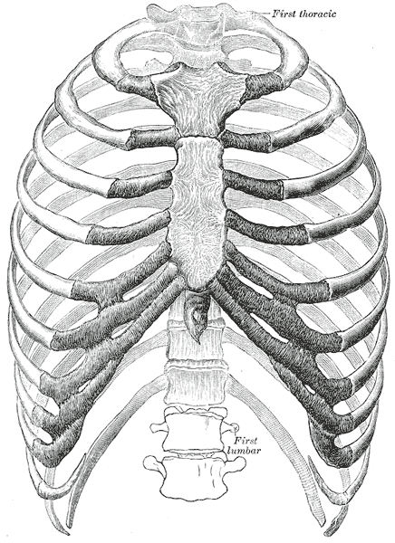
The adult skeleton has 206 bones in two divisions. The axial skeleton — 80 bones — forms the central axis: the skull, the vertebral column of 33 vertebrae in five regions, and the thoracic cage of 12 rib pairs and the sternum. Its main job is protection, and the shapes follow from that. The appendicular skeleton — 126 bones — comprises the limbs and the two girdles that attach them: the pectoral girdle of clavicle and scapula, which is deliberately loose and mobile, and the pelvic girdle, which is fused and stable. That contrast is the whole trade-off of the skeleton in miniature: the shoulder sacrifices stability for reach, the hip sacrifices reach for load-bearing.
Bones are classified by shape and the shape predicts the function. Long bones such as the femur and humerus act as levers, with a shaft and two expanded ends. Short bones such as the carpals and tarsals are roughly cubic and provide stability with limited gliding movement. Flat bones such as the skull vault, sternum, ribs and scapula protect and offer broad muscle attachment. Irregular bones such as the vertebrae and facial bones do jobs too specific for a simple category. Sesamoid bones, of which the patella is the largest, sit within tendons and improve the angle at which a muscle pulls.
A typical long bone has a consistent internal plan. The diaphysis is the shaft, a hollow cylinder of dense cortical bone around a medullary cavity — hollow because a tube resists bending almost as well as a solid rod at a fraction of the weight. The epiphyses are the expanded ends, made of trabecular bone under a shell of cortical bone and capped by articular cartilage where they form a joint. Between them lies the metaphysis, which in a growing child contains the epiphyseal growth plate of hyaline cartilage. The outer surface is covered by periosteum, richly supplied with nerves — which is why a bruised shin hurts so much — and the inner surfaces by endosteum. Both carry the cells that build and remove bone.
How the system works
The skeleton does five things. It supports the body and gives soft tissue something to hang from. It protects: the skull for the brain, the vertebral column for the cord, the ribcage for heart and lungs, the pelvis for the pelvic organs. It enables movement by giving muscles rigid levers and joints to move them about; a muscle can only pull, so movement always requires a bone to pull on and usually an opposing muscle to reverse it. It stores minerals — around 99 per cent of the body's calcium and 85 per cent of its phosphate. And in the red marrow it makes blood, producing all red cells, platelets and white cells.
Two of those jobs conflict, and the resolution is continuous remodelling. About 10 per cent of the adult skeleton is replaced each year by a coupled cycle: osteoclasts, large multinucleate cells derived from the monocyte lineage, seal onto a bone surface and dissolve mineral with acid and matrix with enzymes; osteoblasts then move in and lay down new osteoid which subsequently mineralises. This is what allows bone to respond to load — Wolff's law, that bone is deposited where it is stressed and removed where it is not, which is why weight-bearing exercise builds bone and why astronauts and bed-bound patients lose it rapidly. It is also what makes bone a usable mineral reserve.
Calcium regulation runs through the same machinery and is unusually tight, because blood calcium controls nerve and muscle excitability. When plasma calcium falls, the parathyroid glands release parathyroid hormone, which stimulates osteoclastic resorption, increases calcium reabsorption in the kidney, and activates vitamin D to increase absorption from the gut. When calcium rises, calcitonin from the thyroid opposes it, though its role in adults is minor. Growth is a separate process: long bones lengthen only at the epiphyseal plates, where chondrocytes proliferate on the epiphyseal side and are replaced by bone on the diaphyseal side, until oestrogen closes the plates in late adolescence — in both sexes, which is why disorders of oestrogen signalling affect final height in men too.
The cells that do the work
Four cell types run bone and they are best learned as a sequence. Osteoprogenitor cells in periosteum and endosteum are the stem population. Osteoblasts differentiate from them and build: they secrete osteoid, which is mostly type I collagen with proteoglycans, then control its mineralisation with calcium hydroxyapatite crystals. Osteocytes are osteoblasts that have walled themselves into the matrix, sitting in lacunae and connected to each other through canaliculi by long cell processes; they are not dormant but act as the tissue's mechanical sensors, detecting strain and signalling for remodelling where it is needed. Osteoclasts are the demolition cells, large and multinucleate, derived from the same lineage as macrophages rather than from the other three, and they attach to bone with a ruffled border and secrete acid and cathepsin K into a sealed compartment. The matrix itself explains bone's mechanical behaviour: roughly 35 per cent organic, mostly collagen, which gives tensile strength and flexibility, and 65 per cent mineral, which gives compressive strength and rigidity. Remove the mineral and a bone becomes rubbery; burn away the collagen and it becomes chalk that shatters. Bone needs both, which is why children with brittle collagen fracture easily despite normal mineral, and why elderly bone with normal collagen but reduced mineral fractures too.
Under the microscope
Mature bone is lamellar, its collagen laid down in parallel sheets with alternating fibre orientation in successive layers, which resists force from several directions at once. In compact bone the unit is the osteon, or Haversian system: concentric lamellae arranged around a central Haversian canal carrying vessels and nerves, with osteocytes in lacunae between the lamellae and canaliculi radiating outward like spokes. Volkmann canals run transversely, connecting Haversian canals to each other and to the outer and inner surfaces. Between intact osteons lie interstitial lamellae, the remnants of osteons removed by earlier remodelling cycles, and they are a visible record of turnover. Circumferential lamellae wrap the outer and inner surfaces of the whole shaft. Trabecular bone contains no osteons at all, because its struts are thin enough to be fed by diffusion from marrow, so it is built from parallel lamellae instead. Woven bone, with randomly arranged collagen, is mechanically weaker and is normal only in the fetus and in fracture callus; finding it in adult tissue means something is being laid down too fast.
When the system fails
Osteoporosis is uncoupled remodelling: resorption outpaces formation, bone mass falls and microarchitecture deteriorates, until fractures occur under loads that would not previously have caused them. It is silent until the first fracture, most often of the wrist, vertebra or hip, and it accelerates sharply after the menopause because oestrogen restrains osteoclasts. It is distinct from osteomalacia, and confusing the two leads to the wrong treatment: in osteoporosis there is too little bone of normal composition, whereas in osteomalacia — rickets in children — there is a normal quantity of bone that is inadequately mineralised, almost always from vitamin D deficiency.
Fracture healing runs a predictable timetable and explains why immobilisation works. A haematoma forms in the first hours, followed by an inflammatory phase; a soft callus of fibrocartilage bridges the gap over one to three weeks; that is replaced by a hard callus of woven bone over the following weeks; and remodelling then converts woven bone to lamellar bone and restores the original contour over months to years. Movement at the fracture site disrupts the soft callus, which is why fixation matters, and a poor blood supply is why some sites — the scaphoid, the femoral neck, the talus — are notorious for failing to unite.
Two further categories complete the clinical picture. Osteoarthritis is often listed with the skeleton but is a disease of the joint, beginning in articular cartilage; the bony changes — sclerosis, osteophytes, subchondral cysts — are secondary responses to the loss of the cartilage above. Bone infection, osteomyelitis, is difficult to eradicate because dead bone fragments become sequestra with no blood supply, so antibiotics cannot reach the organisms inside them and surgery is often needed. Finally, bone is one of the commonest sites for metastasis, particularly from breast, prostate, lung, kidney and thyroid, and pathological fracture through a metastasis is sometimes the first sign of the primary cancer.
Common questions about the skeletal system
What are the functions of the skeletal system?
Five. Support, giving the body its frame; protection of the brain, spinal cord, heart, lungs and pelvic organs; movement, by providing rigid levers and joints for muscles to pull on; mineral storage, holding about 99 per cent of the body's calcium and 85 per cent of its phosphate; and blood cell production in the red marrow, which makes all red cells, white cells and platelets.
How many bones are in the human body?
206 in a typical adult — 80 in the axial skeleton (skull, vertebral column, thoracic cage) and 126 in the appendicular skeleton (limbs and girdles). A newborn has around 270, because several bones such as the sacrum, coccyx and the parts of the hip bone start separate and fuse during growth. Small variations exist between individuals, mainly in the number of sesamoid bones.
What is the difference between compact and spongy bone?
Compact, or cortical, bone is dense and forms the outer shell and the shafts of long bones; its unit is the osteon, concentric lamellae around a central canal carrying vessels. Spongy, or trabecular, bone forms a lattice of struts inside the ends of long bones and within flat bones; it has no osteons because the struts are thin enough to be fed by diffusion from the marrow between them. Trabecular bone is lighter, remodels faster, and is where osteoporotic loss shows first.
Is bone alive?
Entirely. It has its own blood supply, four resident cell populations, and it is dismantled and rebuilt continuously at about 10 per cent per year. Osteocytes buried in the matrix act as strain sensors and direct where remodelling happens, which is how bone thickens in response to loading and thins without it. That is also why prolonged bed rest and spaceflight cause rapid bone loss.
Selected references
- Standring S, ed. Gray's Anatomy: The Anatomical Basis of Clinical Practice. 42nd ed. Elsevier; 2021.
- Ross MH, Pawlina W. Histology: A Text and Atlas. 8th ed. Wolters Kluwer; 2020.
- Hall JE, Hall ME. Guyton and Hall Textbook of Medical Physiology. 14th ed. Elsevier; 2021.
- Florencio-Silva R, Sasso GR, Sasso-Cerri E, Simões MJ, Cerri PS. Biology of bone tissue: structure, function, and factors that influence bone cells. BioMed Research International. 2015;2015:421746.
- Raisz LG. Pathogenesis of osteoporosis: concepts, conflicts, and prospects. Journal of Clinical Investigation. 2005;115(12):3318-3325.
- Kumar V, Abbas AK, Aster JC. Robbins and Cotran Pathologic Basis of Disease. 10th ed. Elsevier; 2021.
Medical disclaimer. This article is written for education and general understanding. It is not medical advice and cannot replace assessment by a qualified healthcare professional. If you have symptoms or health concerns, speak with a clinician. Read the full disclaimer.