The cardiovascular system exists to solve one problem: every cell in a body larger than a few millimetres is too far from the outside world to get its own oxygen. Diffusion alone would take days to move a molecule from your lungs to your foot. So the body builds a delivery network instead — a pump, a set of pipes with adjustable diameter, and a fluid that carries seventy times more oxygen than water would. Understanding it means holding two ideas at once: the heart is two pumps in one organ, and almost everything the system does is aimed at keeping the pressure inside it within a narrow range.
The parts, and how they fit together
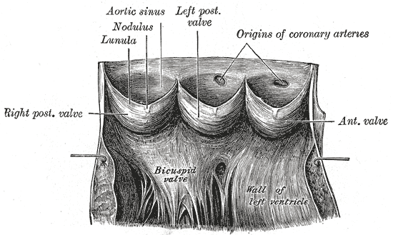
The heart sits in the middle mediastinum and contains four chambers arranged as two pumps side by side. The right atrium and right ventricle take deoxygenated blood returning from the body and send it to the lungs; the left atrium and left ventricle take the oxygenated blood coming back and send it to everywhere else. The two circuits are in series, not parallel, which means every drop of blood alternates between them and the two sides must move exactly the same volume per minute or blood accumulates on one side. The wall of the left ventricle is three to four times thicker than the right, not because it moves more blood but because it works against a far higher pressure.
The vessels form a branching tree with a specific job at each generation. Elastic arteries such as the aorta stretch in systole and recoil in diastole, smoothing the heart's intermittent output into continuous flow. Muscular arteries distribute blood to organs. Arterioles, with only a few layers of smooth muscle, are the resistance vessels, and because resistance varies with the fourth power of radius, small changes in their diameter dominate blood pressure. Capillaries — a single endothelial cell thick and around ten billion of them — are where the whole system finally does its job. Venules and veins collect the blood and return it, holding around two-thirds of the total volume at any moment.
Two circulations run off this arrangement. The pulmonary circulation carries blood from the right ventricle through the pulmonary arteries to the lungs and back through the pulmonary veins, and it operates at roughly a fifth of systemic pressure because the lungs sit immediately adjacent and their capillaries are fragile. The systemic circulation supplies everything else and includes two important special cases: the coronary circulation, which supplies the heart itself and fills during diastole rather than systole, and the hepatic portal system, in which blood from the gut passes through a second capillary bed in the liver before returning to the heart.
How the system works
The pump is electrical before it is mechanical. Pacemaker cells in the sinoatrial node depolarise spontaneously, setting a rate of about 100 per minute that resting vagal tone slows to 60 to 80. The impulse crosses the atria, is delayed roughly 0.1 seconds at the atrioventricular node so atrial contraction can finish filling the ventricles, then runs rapidly down the bundle of His and Purkinje fibres so the ventricles contract from the apex upward. The fibrous skeleton insulates atria from ventricles electrically, making the atrioventricular bundle the only normal route between them — which is why blocking it produces heart block.
Output is the product of two numbers. Cardiac output equals heart rate multiplied by stroke volume, and at rest that is about 70 beats per minute times 70 millilitres, or roughly five litres per minute — the body's entire blood volume every minute. In hard exercise it can reach 20 to 25 litres, and in a trained athlete more. Stroke volume itself depends on three things: preload, the degree of stretch before contraction, described by the Frank-Starling relationship in which a more filled ventricle contracts more forcefully; afterload, the pressure the ventricle must exceed to eject; and contractility, the intrinsic vigour of the muscle, which sympathetic stimulation and circulating adrenaline increase.
Blood pressure is what the system regulates most carefully, because too little means organs are not perfused and too much damages the vessels themselves. Mean arterial pressure is cardiac output multiplied by total peripheral resistance, so the body has exactly two levers. Second by second, baroreceptors in the carotid sinus and aortic arch sense stretch and adjust vagal and sympathetic outflow, changing heart rate, contractility and arteriolar tone within a beat or two — this is why standing up quickly does not usually make you faint. Over hours to days the kidney takes over through the renin-angiotensin-aldosterone system, adjusting sodium and water retention and therefore blood volume. Local control overrides both where it matters: active tissue produces carbon dioxide, adenosine and heat, all of which dilate local arterioles and steer flow toward the organs that need it.
The cells that do the work
Three cell populations carry the system. Cardiomyocytes are short, branched, striated cells joined at intercalated discs, which combine desmosomes and fascia adherens for mechanical continuity with connexin gap junctions for electrical continuity, letting the myocardium behave as one functional syncytium. Up to a third of their volume is mitochondria because the heart is almost entirely aerobic, and they renew at well under one per cent a year, which is why lost muscle is replaced by scar rather than by muscle. Vascular smooth muscle cells set the diameter of every artery and arteriole, contracting in response to sympathetic noradrenaline, angiotensin II and stretch, relaxing in response to nitric oxide and local metabolites. The endothelium, a single continuous cell layer weighing about a kilogram in total, turns out to be the system's most active tissue: it releases nitric oxide and prostacyclin to relax the muscle beneath it, endothelin to constrict it, presents an anticoagulant surface in health and a procoagulant, leukocyte-adhesive one when injured. Almost every cardiovascular disease begins as endothelial dysfunction, which is why the same risk factors — smoking, hypertension, diabetes, high LDL cholesterol — damage arteries in every territory at once.
Under the microscope
The three layers repeat everywhere with different proportions, and reading the proportions tells you what you are looking at. The heart wall has epicardium, a mesothelium over fat and coronary vessels; myocardium, the thick middle layer of branching striated fibres with central nuclei and dark intercalated discs; and endocardium, an endothelium continuous with the vascular lining, sitting on connective tissue that carries the Purkinje fibres. Vessels follow the same plan as tunica intima, media and adventitia. In a muscular artery the media is thick and obviously muscular, bounded internally by a wavy internal elastic lamina whose crinkled appearance is a fixation artefact and is diagnostic. In an elastic artery the media is filled with parallel wavy sheets of elastin. Veins have a thin media and a relatively thick adventitia, so on a slide an artery is round with a small lumen while the vein beside it is collapsed with a wide one. Capillaries have no wall beyond a single endothelial nucleus bulging into a lumen barely wide enough for one red cell, and they come in continuous, fenestrated and sinusoidal forms depending on how much the organ needs to let through.
When the system fails
Atherosclerosis is the dominant disease of the system and is a disease of the arterial intima. Endothelial injury lets LDL into the subendothelial space, macrophages engulf it and become foam cells, smooth muscle migrates in from the media and builds a fibrous cap over a lipid core. The narrowing itself causes symptoms only on exertion; the dangerous event is sudden, when the cap ruptures, exposes thrombogenic material, and a clot occludes the vessel within minutes. The same process produces myocardial infarction in a coronary artery, ischaemic stroke in a cerebral one, and claudication or limb ischaemia in the legs.
Hypertension is the commonest chronic disorder of the system and, because it is silent, the most often missed. Sustained high pressure thickens the left ventricle, which initially preserves output and eventually stiffens the chamber so it cannot fill; it accelerates atherosclerosis; and it damages the small vessels of the kidney, retina and brain. Heart failure is the endpoint of many of these paths: a heart that cannot meet demand at normal filling pressures, either because it contracts poorly or because it fills poorly. The compensations that help acutely — sympathetic drive and renin-angiotensin-aldosterone activation — become harmful over months, which is why most modern heart failure drugs work by blocking precisely those systems.
Failures of rhythm and of the return circuit complete the picture. Atrial fibrillation, the commonest sustained arrhythmia, replaces coordinated atrial contraction with chaos; blood stagnates in the left atrial appendage, clots form, and the resulting embolus commonly lodges in the brain, which is why the treatment is anticoagulation as much as rate control. On the venous side, deep vein thrombosis follows Virchow's triad of stasis, endothelial injury and hypercoagulability, and its danger is a fragment travelling through the right heart to lodge in the pulmonary arteries. Valve disease loads the ventricle in two distinct ways: stenosis imposes a pressure load and drives concentric hypertrophy, regurgitation imposes a volume load and drives dilatation.
Common questions about the cardiovascular system
What does the cardiovascular system do?
It delivers oxygen and nutrients to every cell and removes carbon dioxide and waste, because diffusion alone is far too slow across the distances in a human body. It also distributes hormones and immune cells, spreads heat from the core to the skin, and maintains fluid and pH balance. All of that depends on keeping blood pressure high enough to perfuse organs but low enough not to damage the vessels.
What is the difference between the pulmonary and systemic circulation?
The pulmonary circulation runs from the right ventricle to the lungs and back to the left atrium, picking up oxygen and releasing carbon dioxide; it operates at about a fifth of systemic pressure because the lungs are close by and their capillaries are delicate. The systemic circulation runs from the left ventricle to the rest of the body and back to the right atrium, at much higher pressure. They are in series, so every drop of blood passes through both in turn.
What determines blood pressure?
Mean arterial pressure equals cardiac output multiplied by total peripheral resistance, so the body adjusts either how much blood the heart moves or how tight the arterioles are. Second by second, baroreceptors in the carotid sinus and aortic arch alter heart rate, contractility and vessel tone. Over hours and days the kidney adjusts blood volume through the renin-angiotensin-aldosterone system. Most blood pressure medication works on one of those two levers.
What is cardiac output and what is normal?
Cardiac output is heart rate multiplied by stroke volume — the volume of blood the heart pumps per minute. At rest it is roughly 70 beats per minute times 70 millilitres, about five litres, which is close to the body's entire blood volume every minute. During hard exercise it can rise to 20 to 25 litres per minute, and higher still in trained endurance athletes.
Selected references
- Hall JE, Hall ME. Guyton and Hall Textbook of Medical Physiology. 14th ed. Elsevier; 2021.
- Standring S, ed. Gray's Anatomy: The Anatomical Basis of Clinical Practice. 42nd ed. Elsevier; 2021.
- Klabunde RE. Cardiovascular Physiology Concepts. 3rd ed. Wolters Kluwer; 2021.
- Pober JS, Sessa WC. Evolving functions of endothelial cells in inflammation. Nature Reviews Immunology. 2007;7(10):803-815.
- Libby P, Buring JE, Badimon L, et al. Atherosclerosis. Nature Reviews Disease Primers. 2019;5(1):56.
- Kumar V, Abbas AK, Aster JC. Robbins and Cotran Pathologic Basis of Disease. 10th ed. Elsevier; 2021.
Medical disclaimer. This article is written for education and general understanding. It is not medical advice and cannot replace assessment by a qualified healthcare professional. If you have symptoms or health concerns, speak with a clinician. Read the full disclaimer.