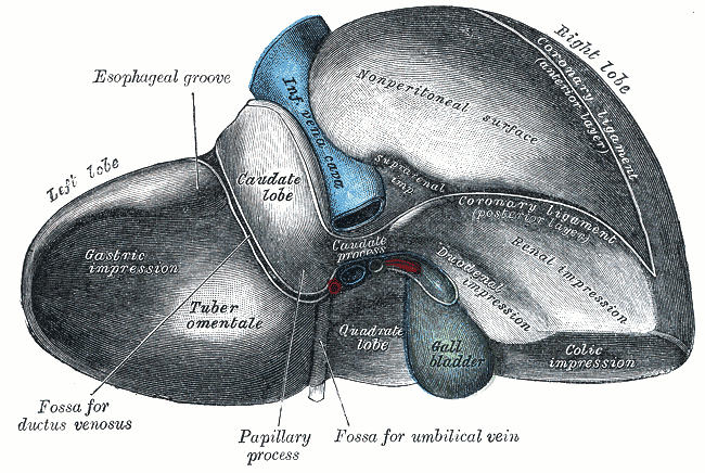
The liver is the largest internal organ and the body's central biochemical processing plant. Weighing around 1.5 kilograms, it receives roughly a quarter of cardiac output, performs several hundred distinct metabolic functions, and is the only major internal organ capable of substantial regeneration. Every nutrient absorbed from the gut passes through it before reaching the rest of the body, which makes it both the first opportunity for processing and the first line of chemical defence.
Anatomy: the structure
The liver occupies the right upper quadrant beneath the diaphragm, protected by the lower ribs and covered almost entirely by visceral peritoneum. Classical anatomy divides it into right and left lobes by the falciform ligament, with the caudate and quadrate lobes on the visceral surface. Surgically, however, the functional division follows Couinaud segmentation: eight independent segments, each with its own portal inflow, hepatic arterial supply, and biliary drainage, which is what makes partial hepatectomy along segmental planes possible.
The dual blood supply is the defining feature of hepatic anatomy. The hepatic artery proper delivers about 25 percent of the blood but the majority of the oxygen, while the portal vein delivers about 75 percent of the blood, nutrient-rich and partly deoxygenated, draining the entire gastrointestinal tract, spleen, and pancreas. Both converge at the hepatic sinusoids, and blood exits through central veins into the hepatic veins and then the inferior vena cava.
Bile flows in the opposite direction to blood. Canaliculi between hepatocytes drain into bile ductules, then into the right and left hepatic ducts, which join as the common hepatic duct. The cystic duct connects the gallbladder, where bile is concentrated up to tenfold, and the resulting common bile duct joins the pancreatic duct at the ampulla of Vater, guarded by the sphincter of Oddi. The portal triad running in the free edge of the lesser omentum carries the portal vein, hepatic artery, and bile duct together.
Physiology: how it works
In carbohydrate metabolism, the liver buffers blood glucose in both directions. After a meal it takes up glucose and stores it as glycogen; between meals it releases glucose by glycogenolysis; and in prolonged fasting it synthesises glucose de novo through gluconeogenesis using lactate, glycerol, and amino acids. In lipid metabolism it oxidises fatty acids, produces ketone bodies during fasting, synthesises cholesterol and lipoproteins, and packages triglycerides into VLDL for export.
In protein metabolism, the liver synthesises almost all plasma proteins, including albumin, which supplies most of plasma oncotic pressure, and the majority of coagulation factors, several of which require vitamin K. It also performs deamination of amino acids and converts the resulting toxic ammonia into urea for renal excretion, which is why liver failure and hyperammonaemia are linked. Serum albumin and prothrombin time therefore serve as practical measures of synthetic liver function.
Detoxification proceeds in two phases. Phase I reactions, driven mainly by cytochrome P450 enzymes, add or expose reactive groups through oxidation, reduction, or hydrolysis. Phase II reactions conjugate the product with glucuronate, sulfate, glutathione, or an amino acid, making it water-soluble for biliary or renal excretion. The liver also conjugates bilirubin from haem breakdown, secretes bile salts that emulsify dietary fat, and stores vitamins A, D, B12, and K along with iron and copper.
Biology: the living cells
Hepatocytes make up around 80 percent of liver volume and are among the most metabolically versatile cells in the body. They are polarised, with a basolateral surface facing the sinusoid for exchange with blood and an apical surface forming the bile canaliculus, and they are packed with smooth endoplasmic reticulum carrying cytochrome P450 enzymes, rough endoplasmic reticulum for protein synthesis, abundant mitochondria, peroxisomes, and glycogen granules. Many are binucleate or polyploid. Three other populations matter: Kupffer cells, resident macrophages lining the sinusoids that clear bacteria and aged erythrocytes arriving in portal blood; hepatic stellate cells in the space of Disse, which store vitamin A in health but transform into collagen-secreting myofibroblasts in chronic injury; and liver sinusoidal endothelial cells, which are fenestrated and lack a continuous basement membrane so that plasma contacts hepatocytes directly.
Histology: under the microscope
Liver tissue can be read through three overlapping models. The classic lobule is a hexagon with a central vein at its centre and portal triads at its corners, with plates of hepatocytes one or two cells thick radiating outward, separated by sinusoids. The portal lobule reorients the same tissue around a bile duct to describe exocrine secretion. The hepatic acinus, most useful clinically, is centred on the vessels between two portal triads and divides parenchyma into three zones: zone 1 nearest the blood supply is oxygen-rich and handles gluconeogenesis, zone 3 nearest the central vein is oxygen-poor, rich in cytochrome P450, and therefore most vulnerable to both ischaemic and toxic injury. Between the sinusoid and hepatocyte lies the space of Disse, containing stellate cells and forming the origin of hepatic lymph.
Clinical correlations
Cirrhosis is the common endpoint of chronic liver injury from alcohol, viral hepatitis, or metabolic dysfunction-associated steatotic liver disease. Activated stellate cells deposit collagen, normal architecture is replaced by regenerative nodules surrounded by fibrous septa, and the consequences follow directly from the anatomy: obstructed sinusoidal flow raises portal pressure, producing varices, splenomegaly, and ascites, while lost hepatocyte mass reduces albumin, clotting factors, and urea synthesis.
Jaundice becomes visible when bilirubin exceeds roughly 2 to 3 mg/dL, and its cause can be localised by the pattern. Prehepatic jaundice from haemolysis raises unconjugated bilirubin; hepatic jaundice from hepatocellular damage raises both fractions; posthepatic jaundice from biliary obstruction raises conjugated bilirubin with pale stools and dark urine. Liver enzyme patterns are similarly informative, with ALT and AST indicating hepatocellular injury and ALP with GGT indicating cholestasis.
The first-pass effect has direct pharmacological consequences: drugs absorbed from the gut reach the liver through the portal vein before entering the systemic circulation, so a substantial fraction may be metabolised before it ever acts. Paracetamol overdose illustrates the limits of this system, since once glucuronidation and sulfation are saturated, the reactive metabolite NAPQI depletes glutathione and causes centrilobular zone 3 necrosis, which is why N-acetylcysteine given early is effective.
Common questions about the liver
Why does the liver have two separate blood supplies?
The hepatic artery delivers oxygenated blood at systemic pressure to meet the organ's metabolic needs, while the portal vein delivers nutrient-rich blood collected from the intestines, spleen, and pancreas. This arrangement lets the liver process everything absorbed from the gut before it reaches the general circulation.
What is a portal triad?
A portal triad sits at the corner of each classic liver lobule and contains a branch of the portal vein, a branch of the hepatic artery, and a bile duct, usually accompanied by lymphatics and nerves. Blood flows from the triad through sinusoids toward the central vein, while bile flows in the opposite direction toward the duct.
How does the liver detoxify drugs and toxins?
Phase I reactions, mainly by cytochrome P450 enzymes, oxidise or otherwise modify the compound to expose a reactive group. Phase II reactions then conjugate that product with molecules such as glucuronate, sulfate, or glutathione, making it water-soluble so it can be excreted in bile or urine.
Can the liver really grow back?
Yes, within limits. Remaining hepatocytes re-enter the cell cycle and restore liver mass after partial resection, typically within weeks. This works when the underlying framework is intact, but in cirrhosis the scarred architecture prevents functional regeneration even though nodules of new cells form.
Selected references
- Standring S, ed. Gray's Anatomy: The Anatomical Basis of Clinical Practice. 42nd ed. Elsevier; 2021.
- Hall JE, Hall ME. Guyton and Hall Textbook of Medical Physiology. 14th ed. Elsevier; 2021.
- Ross MH, Pawlina W. Histology: A Text and Atlas. 8th ed. Wolters Kluwer; 2020.
- Rappaport AM. The microcirculatory hepatic unit. Microvascular Research. 1973;6(2):212-228.
- Boron WF, Boulpaep EL. Medical Physiology. 3rd ed. Elsevier; 2017.
- Kumar V, Abbas AK, Aster JC. Robbins and Cotran Pathologic Basis of Disease. 10th ed. Elsevier; 2021.
Medical disclaimer. This article is written for education and general understanding. It is not medical advice and cannot replace assessment by a qualified healthcare professional. If you have symptoms or health concerns, speak with a clinician. Read the full disclaimer.