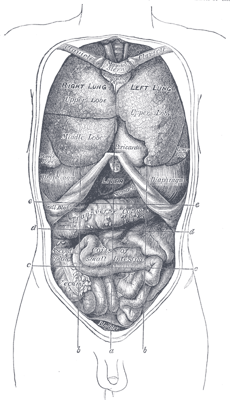
The digestive tract is, topologically, the outside of your body threaded through the middle of it. Food in the lumen has not entered you until it crosses an epithelial cell, and almost everything the system does is aimed at that one crossing: breaking large insoluble molecules into small soluble ones, moving them along at the right pace, and presenting them to an absorptive surface that is folded and refolded until nine metres of tube offers a surface area measured in tens of square metres. Four accessory organs — salivary glands, liver, gallbladder and pancreas — never touch the food but supply almost all the chemistry.
The parts, and how they fit together
The tract runs continuously from mouth to anus and each station does something distinct. The mouth mechanically breaks food and mixes it with saliva; the pharynx and oesophagus transport it, the latter through a 25-centimetre muscular tube whose upper third is skeletal muscle and lower third smooth, with sphincters at each end. The stomach is a reservoir and a mixer, holding up to about 1.5 litres, churning food into semi-liquid chyme and beginning protein digestion in acid. The small intestine — duodenum, jejunum, ileum, around six metres — is where digestion is completed and where over 90 per cent of absorption happens. The large intestine, about 1.5 metres, recovers water and electrolytes and houses the microbiota.
The four accessory organs supply the chemistry. Salivary glands produce a litre or more of saliva daily, containing amylase, lubricating mucus and antibacterial lysozyme. The liver manufactures bile, which contains no enzymes at all but supplies bile salts that emulsify fat into droplets small enough for enzymes to work on. The gallbladder stores and concentrates that bile between meals and contracts when fat reaches the duodenum. The pancreas is the workhorse, secreting bicarbonate to neutralise gastric acid and a full set of enzymes — amylase, lipase, and proteases secreted as inactive precursors so they do not digest the pancreas itself.
From oesophagus to anus the wall keeps the same four layers, which is one of the most useful facts in gastrointestinal medicine. The mucosa is the innermost, comprising epithelium, a loose connective tissue lamina propria and a thin muscularis mucosae. The submucosa is denser connective tissue carrying larger vessels and the submucosal nerve plexus. The muscularis externa is usually an inner circular and an outer longitudinal layer of smooth muscle with the myenteric plexus between them. The outermost layer is serosa where the organ is intraperitoneal and adventitia where it is retroperitoneal. Regional specialisations — the stomach's extra oblique muscle layer, the colon's taeniae coli — are variations on that theme.
How the system works
Digestion is a relay of enzymes, each working in the conditions the previous station created. Salivary amylase starts on starch and is then destroyed by gastric acid. In the stomach, parietal cells secrete hydrochloric acid that brings pH to around 1.5 to 3.5, denaturing protein and converting pepsinogen from chief cells into active pepsin; acid also kills most swallowed bacteria and frees dietary iron and vitamin B12. In the duodenum, pancreatic bicarbonate raises pH toward 7 to 8, which is where pancreatic enzymes work: amylase finishes carbohydrate, trypsin and chymotrypsin continue protein, and lipase, helped by bile salt emulsification, handles fat. Final digestion happens at the brush border itself, where enzymes such as lactase and maltase are embedded in the microvillous membrane — which is why lactose intolerance is a deficiency of a membrane enzyme rather than a secreted one.
Absorption is a problem of surface area, solved three times over. The intestinal lining is thrown into circular folds, each fold is covered in finger-like villi about a millimetre tall, and each absorptive cell carries thousands of microvilli forming a brush border. Together these multiply the plain tube's surface by several hundredfold. Sugars and amino acids are taken up by sodium-coupled transporters and pass into capillaries draining to the hepatic portal vein, so everything absorbed goes through the liver before reaching the rest of the body. Fat takes a different route: digested into fatty acids and monoglycerides, carried in bile salt micelles to the cell surface, reassembled inside the cell into triglycerides, packaged as chylomicrons, and released into the lacteal, a lymphatic vessel in the centre of each villus, bypassing the liver entirely.
Movement and secretion are coordinated by a nervous system the gut keeps for itself. The enteric nervous system contains several hundred million neurons in the myenteric and submucosal plexuses and can run reflexes with no input from the brain, which is why an isolated segment of intestine still performs peristalsis. Its interstitial cells of Cajal act as pacemakers, generating slow waves that set the rhythm. Peristalsis propels contents; segmentation mixes them. The autonomic nervous system modulates all of it, parasympathetic activity increasing motility and secretion and sympathetic reducing them. Layered on top is hormonal control from enteroendocrine cells in the gut lining: gastrin stimulates acid, secretin triggers pancreatic bicarbonate, cholecystokinin contracts the gallbladder and releases pancreatic enzymes, and hormones including GLP-1 signal satiety to the brain.
The cells that do the work
The intestinal epithelium is the fastest-renewing tissue in the body and the arrangement that allows it is elegant. Stem cells sit at the base of the crypts of Lieberkühn, dividing continuously; their progeny migrate up the villus, mature into absorptive enterocytes, work for a few days at the tip and are shed into the lumen, the entire lining being replaced every three to five days. Four differentiated cell types come off that production line. Enterocytes, with their brush border and embedded digestive enzymes, do the absorbing. Goblet cells secrete protective mucus and become steadily more numerous toward the colon. Enteroendocrine cells release the hormones above. Paneth cells stay at the crypt base and secrete lysozyme and defensins, protecting the stem cells beneath them from bacteria. The stomach has its own set: parietal cells packed with mitochondria and an internal membrane system that everts into a secretory canaliculus when acid production begins, chief cells full of zymogen granules, and mucous neck cells producing the bicarbonate-rich mucus layer that stops the stomach digesting itself. Alongside all of this lives the microbiota, on the order of a hundred trillion organisms concentrated in the colon, which ferment indigestible fibre into short-chain fatty acids that colonocytes use as their main fuel, synthesise vitamin K and several B vitamins, and compete with pathogens for space.
Under the microscope
Each region announces itself by its mucosa. The oesophagus is lined by non-keratinised stratified squamous epithelium for abrasion resistance, changing abruptly at the gastro-oesophageal junction — the Z line — to the simple columnar epithelium of the stomach. Gastric mucosa is a field of closely packed pits leading into glands, and the cell mix identifies the region: the fundus and body are full of pink parietal cells and basophilic chief cells, the pylorus is mostly mucus-secreting with gastrin cells. The duodenum is recognised instantly by Brunner's glands in the submucosa, found nowhere else, which secrete alkaline mucus. The jejunum has the tallest, most closely packed villi and no distinguishing extras, which is itself the clue. The ileum carries Peyer's patches, large aggregates of lymphoid tissue in the wall. The colon has no villi at all — a flat surface with straight, deep, test-tube-like crypts and abundant goblet cells, which together are diagnostic. The appendix is the same as the colon but with a small irregular lumen and lymphoid tissue filling the wall.
When the system fails
Failures of the mucosal barrier and its acid balance dominate the upper tract. Gastro-oesophageal reflux disease lets acid into an oesophagus with squamous epithelium not built for it; chronic exposure can convert the lining to intestinal-type columnar epithelium, which is Barrett's oesophagus and carries a raised cancer risk. Peptic ulcer disease is a breach of the mucosa, and the great majority are caused by Helicobacter pylori infection or by NSAIDs, both of which undermine the protective mucus and bicarbonate layer rather than raising acid dramatically. Recognising this changed treatment from acid suppression alone to eradicating the organism.
Malabsorption presents as symptoms out of proportion to appetite. In coeliac disease, an immune response to gluten flattens the villi and destroys the absorptive surface, producing diarrhoea, weight loss, anaemia and fatigue; because the damage is proximal, iron and folate deficiency come first. Lactose intolerance is far simpler, being loss of a single brush border enzyme, leaving lactose in the lumen to be fermented by bacteria. Crohn's disease and ulcerative colitis are the two inflammatory bowel diseases and differ in a way that matters: Crohn's can affect any part of the tract with skip lesions and full-thickness inflammation, while ulcerative colitis is continuous, starts at the rectum and involves only the mucosa.
Two surgical patterns are worth carrying. Appendicitis begins when the appendiceal lumen obstructs, and its pain classically starts as a poorly localised central ache referred from the midgut, then shifts to the right iliac fossa once the inflamed appendix irritates the parietal peritoneum — a textbook demonstration of visceral versus somatic pain. Bowel obstruction, whether from adhesions, hernia or tumour, produces colicky pain, vomiting, distension and absolute constipation. Colorectal cancer follows a well-mapped adenoma-to-carcinoma sequence over years, which is precisely why screening colonoscopy works: it removes the adenoma before the sequence completes.
Common questions about the digestive system
What are the organs of the digestive system?
The tract itself runs mouth, pharynx, oesophagus, stomach, small intestine (duodenum, jejunum, ileum), large intestine, rectum and anus. Four accessory organs contribute without food passing through them: the salivary glands, the liver, the gallbladder and the pancreas. The accessory organs supply nearly all the digestive chemistry — bile salts to emulsify fat, and the full enzyme set plus bicarbonate from the pancreas.
Where does most digestion and absorption happen?
In the small intestine, and mostly in the duodenum and jejunum. Pancreatic enzymes finish breaking down carbohydrate, protein and fat there, brush border enzymes complete the final step at the cell membrane, and over 90 per cent of nutrient absorption occurs before the contents reach the large intestine. The colon's job is mainly recovering water and electrolytes and fermenting the fibre that reaches it.
What are the four layers of the gut wall?
From the lumen outward: mucosa, comprising epithelium, lamina propria and muscularis mucosae; submucosa, denser connective tissue carrying vessels and the submucosal nerve plexus; muscularis externa, usually inner circular and outer longitudinal smooth muscle with the myenteric plexus between them; and serosa or adventitia outermost. The plan is the same from oesophagus to anus, with regional variations such as the stomach's third oblique muscle layer.
Why doesn't the stomach digest itself?
Several defences at once. A thick layer of bicarbonate-rich mucus holds a near-neutral pH right at the cell surface even when the lumen is at pH 2. The epithelium has tight junctions preventing acid leaking between cells, and it is replaced every few days. Pepsin is secreted as inactive pepsinogen and only activated once it is out in the acid lumen. Ulcers form when these defences are undermined, usually by Helicobacter pylori or NSAIDs.
Selected references
- Hall JE, Hall ME. Guyton and Hall Textbook of Medical Physiology. 14th ed. Elsevier; 2021.
- Standring S, ed. Gray's Anatomy: The Anatomical Basis of Clinical Practice. 42nd ed. Elsevier; 2021.
- Ross MH, Pawlina W. Histology: A Text and Atlas. 8th ed. Wolters Kluwer; 2020.
- Furness JB. The enteric nervous system and neurogastroenterology. Nature Reviews Gastroenterology and Hepatology. 2012;9(5):286-294.
- Clevers H. The intestinal crypt, a prototype stem cell compartment. Cell. 2013;154(2):274-284.
- Kumar V, Abbas AK, Aster JC. Robbins and Cotran Pathologic Basis of Disease. 10th ed. Elsevier; 2021.
Medical disclaimer. This article is written for education and general understanding. It is not medical advice and cannot replace assessment by a qualified healthcare professional. If you have symptoms or health concerns, speak with a clinician. Read the full disclaimer.