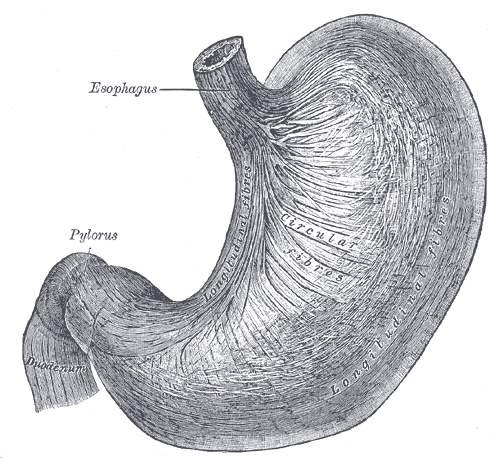
The stomach solves a difficult engineering problem. It must store a meal arriving in minutes, break it down with acid strong enough to reach pH 1 or 2, mix it mechanically into a semi-fluid chyme, release it downstream at a rate the small intestine can handle, and do all of this without digesting its own wall. Each of those requirements is met by a specific anatomical or cellular adaptation.
Anatomy: the structure
The stomach lies mainly in the left upper quadrant, continuous with the oesophagus at the cardia and with the duodenum at the pylorus. It is described in five regions: the cardia surrounding the oesophageal opening, the dome-shaped fundus that sits under the left diaphragm and collects swallowed gas, the large central body, the funnel-shaped pyloric antrum, and the pyloric canal ending at the thick pyloric sphincter. The lesser curvature runs along the right margin and the greater curvature along the left, giving attachment to the lesser and greater omenta.
The wall follows the general gut plan but with one addition. Mucosa, submucosa, muscularis externa, and serosa are all present, but the muscularis externa has three layers rather than two: an inner oblique layer unique to the stomach, a middle circular layer that thickens into the pyloric sphincter, and an outer longitudinal layer. That third layer is what allows genuine churning rather than simple propulsion. When empty, the mucosa and submucosa fold into longitudinal ridges called rugae that flatten as the stomach fills, allowing capacity to rise from about 50 millilitres to 1.5 litres or more.
Blood supply comes from all three branches of the coeliac trunk. The left gastric artery arises directly from the coeliac trunk, the right gastric artery from the hepatic artery, and the gastroepiploic arteries from the hepatic and splenic arteries, forming anastomotic arcades along both curvatures. Venous blood drains into the portal system. Parasympathetic innervation from the vagus nerve stimulates secretion and motility, while sympathetic fibres from the coeliac plexus have the opposite effect, and this is why vagotomy was historically used to reduce acid output.
Physiology: how it works
Acid secretion is the stomach's signature function. Parietal cells use the hydrogen-potassium ATPase, the proton pump, to move protons into the lumen against a concentration gradient of more than a millionfold, generating a luminal pH near 1 to 2. Carbonic anhydrase supplies the protons from carbon dioxide and water, and the bicarbonate produced leaves through the basolateral membrane, causing the postprandial alkaline tide in venous blood. Three stimuli converge on the parietal cell: acetylcholine from vagal fibres, gastrin from antral G cells, and histamine from enterochromaffin-like cells, with histamine acting as the final common amplifier, which is why H2 blockers and proton pump inhibitors work at different points in the same pathway.
Secretion is regulated in three phases. The cephalic phase, triggered by sight, smell, taste, and thought of food, is vagally mediated and accounts for roughly 30 percent of the response before food arrives. The gastric phase, triggered by distension and by peptides in the lumen, accounts for about 60 percent and works largely through gastrin release. The intestinal phase begins as chyme enters the duodenum and is predominantly inhibitory, using secretin, cholecystokinin, and the enterogastric reflex to slow gastric emptying until the duodenum has neutralised and processed what it has received.
Digestion in the stomach is partial rather than complete. Pepsinogen released by chief cells is cleaved to active pepsin by acid, and pepsin begins protein digestion by cutting internal peptide bonds; it is irreversibly inactivated once it reaches the alkaline duodenum. Gastric lipase contributes modestly to fat digestion. Acid also denatures proteins, kills most ingested microorganisms, and converts dietary ferric iron to the absorbable ferrous form. Intrinsic factor, also from parietal cells, is the one truly essential gastric product, since without it vitamin B12 cannot be absorbed in the terminal ileum.
Biology: the living cells
Gastric glands contain a coordinated set of cell types whose proportions change by region. Surface mucous cells cover the entire lining and secrete a thick alkaline mucus. Mucous neck cells sit in the gland neck and secrete a more soluble mucus. Parietal cells, found mainly in the fundus and body, are large eosinophilic cells with abundant mitochondria and an elaborate intracellular canalicular system studded with tubulovesicles that fuse with the membrane when the cell is stimulated, multiplying its secretory surface. Chief cells, concentrated at the gland base, are basophilic with prominent rough endoplasmic reticulum and apical zymogen granules of pepsinogen. Enteroendocrine cells scattered through the epithelium include G cells producing gastrin, mostly in the antrum, D cells producing somatostatin, and enterochromaffin-like cells producing histamine. Stem cells in the isthmus divide continuously and replace the surface epithelium roughly every three to six days.
Histology: under the microscope
The gastric mucosa is lined throughout by simple columnar epithelium that dips into gastric pits, and the ratio of pit depth to gland length is the quickest way to identify the region under the microscope. In the cardia, pits are moderately deep and glands are mainly mucous. In the fundus and body, pits are relatively shallow and the long straight glands are dominated by parietal and chief cells, giving a characteristic two-tone appearance with pink parietal cells higher up and blue chief cells at the base. In the pylorus, pits are deep, glands are short, coiled, and mucous, and G cells are abundant. Beneath the mucosa, the muscularis mucosae separates it from the submucosa, which carries the Meissner plexus, while the Auerbach myenteric plexus sits between the circular and longitudinal muscle layers.
Clinical correlations
Peptic ulcer disease is a failure of the balance between aggressive factors and mucosal defence. Helicobacter pylori infection is the leading cause; the organism survives in the mucus layer by producing urease, which generates ammonia to neutralise local acid, and the resulting chronic inflammation disrupts the barrier. Non-steroidal anti-inflammatory drugs are the second major cause, because inhibiting cyclooxygenase reduces the prostaglandins that maintain mucus, bicarbonate, and mucosal blood flow.
Loss of parietal cells has two separate consequences. Autoimmune atrophic gastritis destroys them, and the resulting intrinsic factor deficiency produces pernicious anaemia with megaloblastic changes and, if untreated, subacute combined degeneration of the spinal cord. The same loss removes acid, so the antrum increases gastrin output, and the sustained hypergastrinaemia can drive enterochromaffin-like cell hyperplasia.
Chronic infection and inflammation also drive a recognised sequence toward malignancy, in which chronic gastritis leads to atrophy, then intestinal metaplasia, then dysplasia, and potentially to gastric adenocarcinoma. Gastro-oesophageal reflux disease reflects a different mechanical problem, incompetence of the lower oesophageal sphincter, where the squamous oesophageal epithelium has none of the stomach's defences and repeated exposure can drive metaplasia to a columnar Barrett epithelium.
Common questions about the stomach
Why does the stomach not digest itself?
The gastric mucosal barrier combines a thick layer of adherent mucus, bicarbonate secreted underneath it that keeps the epithelial surface near neutral pH, tight junctions between epithelial cells, rapid replacement of the surface lining every few days, and generous mucosal blood flow that removes any acid that does penetrate.
What do parietal cells and chief cells each produce?
Parietal cells secrete hydrochloric acid through the proton pump and intrinsic factor, which is required for vitamin B12 absorption in the ileum. Chief cells secrete pepsinogen, the inactive precursor that gastric acid converts into pepsin so protein digestion can begin.
How do proton pump inhibitors reduce stomach acid?
They irreversibly inhibit the hydrogen-potassium ATPase on the apical membrane of parietal cells, which is the final step in acid secretion. Because they act at that final common point rather than on one of the three stimulating signals, they suppress acid more completely than H2 receptor blockers do.
How does Helicobacter pylori survive in stomach acid?
It burrows into the protective mucus layer where the pH is close to neutral, and it produces urease, which splits urea into ammonia and carbon dioxide to buffer the acid immediately around it. The chronic inflammation this causes weakens the mucosal barrier and predisposes to ulceration.
Selected references
- Standring S, ed. Gray's Anatomy: The Anatomical Basis of Clinical Practice. 42nd ed. Elsevier; 2021.
- Hall JE, Hall ME. Guyton and Hall Textbook of Medical Physiology. 14th ed. Elsevier; 2021.
- Ross MH, Pawlina W. Histology: A Text and Atlas. 8th ed. Wolters Kluwer; 2020.
- Barrett KE. Gastrointestinal Physiology. 2nd ed. McGraw Hill; 2014.
- Correa P. Human gastric carcinogenesis: a multistep and multifactorial process. Cancer Research. 1992;52(24):6735-6740.
- Kumar V, Abbas AK, Aster JC. Robbins and Cotran Pathologic Basis of Disease. 10th ed. Elsevier; 2021.
Medical disclaimer. This article is written for education and general understanding. It is not medical advice and cannot replace assessment by a qualified healthcare professional. If you have symptoms or health concerns, speak with a clinician. Read the full disclaimer.