The intestines convert food into the molecules the body actually runs on. The small intestine is where almost all nutrient absorption happens, and its design is an exercise in surface amplification: three successive levels of folding multiply the flat cylindrical area roughly six hundredfold, producing an absorptive surface of around 200 square metres. The large intestine that follows recovers water and electrolytes and houses a microbial ecosystem with more cells than the rest of the body.
Anatomy: the structure
The small intestine runs about 6 metres in three parts. The duodenum, roughly 25 centimetres and mostly retroperitoneal, curves around the head of the pancreas and receives bile and pancreatic juice at the ampulla of Vater. The jejunum, about two fifths of the remaining length, has a thicker wall, larger circular folds, longer vasa recta, and fewer arterial arcades. The ileum, the remaining three fifths, has a thinner wall, more arcades, and abundant Peyer patches, and it ends at the ileocaecal valve.
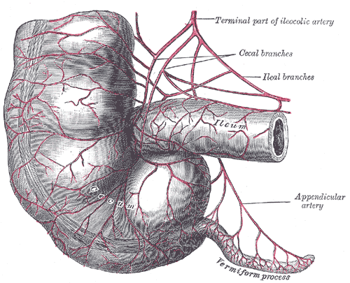
The large intestine is about 1.5 metres and comprises caecum with the appendix, ascending colon, transverse colon, descending colon, sigmoid colon, rectum, and anal canal. Three features distinguish it externally: the taeniae coli, three longitudinal muscle bands that are shorter than the colon itself; the haustra, the sacculations that result; and the omental appendices of fat. Ascending and descending colon are retroperitoneal, while transverse and sigmoid colon hang on mesenteries.
Blood supply follows embryological origin, which explains the watershed zones. The coeliac trunk supplies the foregut as far as the mid-duodenum, the superior mesenteric artery supplies the midgut from there to the proximal two thirds of the transverse colon, and the inferior mesenteric artery supplies the hindgut to the upper rectum. The junction between superior and inferior mesenteric territories near the splenic flexure is a watershed area vulnerable to ischaemic colitis. Venous drainage is portal, so absorbed nutrients pass to the liver first, while lymph from the small intestine carries absorbed fat as chyle to the cisterna chyli and thoracic duct.
Physiology: how it works
Absorptive surface is built in three tiers. Plicae circulares, permanent circular folds of mucosa and submucosa, multiply area about threefold. Villi, finger-like mucosal projections roughly one millimetre tall, multiply it about tenfold more. Microvilli on the apical surface of each enterocyte, forming the brush border, multiply it a further twentyfold. Each villus contains a capillary network for water-soluble nutrients and a central lacteal for absorbed fat.
Digestion finishes at the brush border. Pancreatic amylase leaves carbohydrates as disaccharides, which membrane-bound lactase, sucrase-isomaltase, and maltase split into monosaccharides; glucose and galactose enter through SGLT1 by secondary active transport with sodium, fructose through GLUT5. Peptidases reduce peptides to amino acids and small peptides taken up by PepT1. Fats are emulsified by bile salts into micelles, absorbed as free fatty acids and monoglycerides, reassembled inside the enterocyte, and exported as chylomicrons into the lacteal rather than into blood. Iron is absorbed in the duodenum, folate in the jejunum, and vitamin B12 with bile salts in the terminal ileum.
Motility uses two distinct patterns. Segmentation consists of rhythmic non-propulsive contractions of the circular muscle that mix chyme with enzymes and repeatedly bring it into contact with the mucosa. Peristalsis is a propulsive wave of contraction behind and relaxation ahead of the bolus, coordinated by the enteric nervous system. Between meals the migrating motor complex sweeps residue through every ninety minutes or so. In the colon, mass movements occur only a few times a day, while the colon recovers 1 to 2 litres of water daily and bacterial fermentation of dietary fibre generates short-chain fatty acids that colonocytes use as their preferred fuel.
Biology: the living cells
The intestinal epithelium is the most rapidly renewing tissue in the body, replaced every three to five days from stem cells at the base of the crypts of Lieberkuhn. Those LGR5-positive stem cells give rise to four principal lineages. Enterocytes, the majority, carry the brush border and its digestive enzymes and transporters. Goblet cells secrete mucins, becoming progressively more numerous from duodenum to colon, where mucus protection matters most. Enteroendocrine cells release cholecystokinin, secretin, GIP, GLP-1, and motilin in response to luminal contents, coordinating digestion with the pancreas, gallbladder, and appetite centres. Paneth cells, unique to the small intestine and found at the crypt base, secrete lysozyme, defensins, and phospholipase A2 to control the microbial population near the stem cell niche. Alongside these, the gut-associated lymphoid tissue, including Peyer patches and their overlying M cells, samples luminal antigen, and the colonic microbiome synthesises vitamin K and several B vitamins while competing with potential pathogens.
Histology: under the microscope
The two regions are easy to distinguish under the microscope by asking one question: are there villi? The small intestine has villi with crypts between them; the colon has no villi but straight, closely packed, deep crypts rich in goblet cells. Within the small intestine, the duodenum is identified by Brunner glands in the submucosa, which secrete alkaline mucus to protect against arriving gastric acid. The jejunum shows tall villi and prominent plicae circulares with few lymphoid aggregates. The ileum shows shorter villi, abundant goblet cells, and large Peyer patches in the submucosa. Throughout, the wall follows the four-layer plan of mucosa with its own muscularis mucosae, submucosa carrying the Meissner plexus, muscularis externa with the Auerbach plexus between its circular and longitudinal layers, and serosa or adventitia. In the anal canal, the epithelium changes from simple columnar to stratified squamous at the pectinate line, which also marks the boundary between visceral and somatic innervation.
Clinical correlations
Coeliac disease is an immune reaction to gluten in genetically susceptible people that damages the small intestinal mucosa. The histology is characteristic and mirrors the physiology: villous atrophy, crypt hyperplasia, and intraepithelial lymphocytosis, chiefly in the proximal small intestine. Losing villous surface causes malabsorption, and because iron and folate are absorbed proximally, anaemia is a common presenting feature.
Inflammatory bowel disease has two patterns with different anatomy. Crohn disease can affect any part of the tract from mouth to anus, is transmural, produces skip lesions, non-caseating granulomas, fistulas, and strictures, and most often involves the terminal ileum. Ulcerative colitis is limited to the colon, begins at the rectum and extends continuously, and is confined to mucosa and submucosa, which is why it causes bloody diarrhoea and pseudopolyps rather than fistulas.
Loss of specific segments causes predictable deficits. Terminal ileal resection or disease impairs vitamin B12 and bile salt absorption, causing both anaemia and fat malabsorption. Colorectal cancer typically follows the adenoma to carcinoma sequence over years, which is what makes screening and polyp removal effective. Appendicitis illustrates a purely anatomical mechanism: luminal obstruction raises intraluminal pressure, compromises venous drainage, and leads to ischaemia, bacterial invasion, and perforation if untreated.
Common questions about the intestines
What is the difference between villi and microvilli?
Villi are finger-like projections of the whole mucosa, about one millimetre tall, each containing capillaries and a lacteal. Microvilli are far smaller projections of the apical membrane of a single enterocyte, forming the brush border that carries digestive enzymes and transport proteins. Both increase surface area, but at very different scales.
How can you tell small intestine from large intestine under the microscope?
The presence of villi is the deciding feature. Small intestine has villi projecting into the lumen with crypts between them, while the colon has a flat surface with straight, deep, closely packed crypts and a much higher proportion of goblet cells.
Where are specific nutrients absorbed?
Iron and calcium are absorbed mainly in the duodenum, most carbohydrates, proteins, fats, and folate in the jejunum, and bile salts and vitamin B12 in the terminal ileum. The colon absorbs water, electrolytes, and vitamins produced by bacterial fermentation.
What does the gut microbiome do?
Colonic bacteria ferment fibre the human enzymes cannot digest, producing short-chain fatty acids that nourish the colonic lining. They also synthesise vitamin K and several B vitamins, occupy niches that would otherwise be available to pathogens, and help train the mucosal immune system.
Selected references
- Standring S, ed. Gray's Anatomy: The Anatomical Basis of Clinical Practice. 42nd ed. Elsevier; 2021.
- Barrett KE. Gastrointestinal Physiology. 2nd ed. McGraw Hill; 2014.
- Ross MH, Pawlina W. Histology: A Text and Atlas. 8th ed. Wolters Kluwer; 2020.
- Helander HF, Fandriks L. Surface area of the digestive tract revisited. Scandinavian Journal of Gastroenterology. 2014;49(6):681-689.
- Barker N. Adult intestinal stem cells: critical drivers of epithelial homeostasis and regeneration. Nature Reviews Molecular Cell Biology. 2014;15(1):19-33.
- Hall JE, Hall ME. Guyton and Hall Textbook of Medical Physiology. 14th ed. Elsevier; 2021.
Medical disclaimer. This article is written for education and general understanding. It is not medical advice and cannot replace assessment by a qualified healthcare professional. If you have symptoms or health concerns, speak with a clinician. Read the full disclaimer.