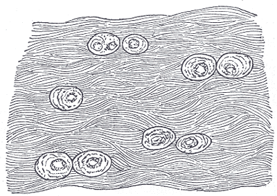
Cartilage is the compromise tissue. It is far stiffer than anything else made of cells and matrix alone, but it bends where bone would break, and it is smooth enough that two cartilage surfaces sliding over each other have less friction than ice on ice. The price of all three properties is the same: the matrix is too dense for blood vessels to grow through. Every chondrocyte in your body is fed by diffusion, and that one fact explains why cartilage grows slowly, heals badly, and, once damaged in a joint, tends to stay damaged.
Where it is, and what it looks like
Hyaline cartilage is the commonest of the three and the default when the word cartilage is used without qualification. It covers the ends of bones inside synovial joints as articular cartilage, forms the costal cartilages joining ribs to sternum, holds the airway open in the rings of the trachea and bronchi, and shapes the nose and larynx. It is also the entire fetal skeleton, later replaced by bone, and it persists at the growth plates of long bones until adolescence. Its matrix is type II collagen in fibrils too fine to see, which is why it looks glassy — hyaline means glass-like.
Elastic cartilage is found in exactly the places that have to bend repeatedly and spring back to an exact shape: the external ear, the epiglottis, the walls of the auditory tube, and parts of the larynx. Its matrix contains the same type II collagen with a dense network of elastic fibres woven through it, which is what gives it flexibility with perfect recoil and, incidentally, a yellowish colour in the fresh state. It is the only one of the three that never calcifies with age.
Fibrocartilage is the toughest and sits wherever a joint has to absorb heavy compression or resist shear. It forms the intervertebral discs, the menisci of the knee, the labra deepening the hip and shoulder sockets, and the pubic symphysis. Structurally it is a hybrid: thick visible bundles of type I collagen, as in tendon, with chondrocytes scattered between them in rows. It has no perichondrium, which is the fibrous sleeve that covers the other two, and it is always found blending into the dense connective tissue or hyaline cartilage around it rather than as a discrete structure.
What it does
Cartilage resists compression through water rather than through solid structure. Its ground substance is packed with aggrecan, a proteoglycan carrying hundreds of negatively charged glycosaminoglycan chains that attract and hold water. Load a joint and water is squeezed out of the matrix into the joint space; unload it and the fixed negative charges draw the water back in. That movement is what cushions the impact, and it is also how the tissue is fed, since with no blood supply the only way nutrients reach a chondrocyte is by being pumped in and out with that water. Cartilage is, quite literally, kept alive by being used.
Articular cartilage adds an extraordinary bearing surface to that. A layer only two to four millimetres thick, lubricated by synovial fluid containing hyaluronan and lubricin, gives a coefficient of friction of around 0.001 to 0.01 — lower than ice sliding on ice, and an order of magnitude better than any engineered bearing. The collagen fibres within it are arranged in arcades, running parallel to the surface at the top, obliquely in the middle and perpendicular to the bone at the base, an architecture that resists shear at the surface while anchoring the tissue firmly below.
Cartilage also grows in two distinct ways, and only one of them survives into adult life. Appositional growth adds new cartilage at the surface, as cells in the inner layer of the perichondrium differentiate into chondroblasts and lay down matrix on the outside. Interstitial growth expands the tissue from within, as chondrocytes already trapped in lacunae divide and secrete new matrix, pushing their neighbours apart. Interstitial growth is what lengthens the epiphyseal growth plates during childhood and stops when they fuse; after that, cartilage in an adult joint can essentially only be maintained, not extended.
The cells that build it
The only cell in cartilage is the chondrocyte, and it lives in a small cavity in the matrix called a lacuna. Chondroblasts, derived from mesenchyme or from the perichondrium, secrete matrix until they have walled themselves in and become chondrocytes; because they often divide once or twice after being trapped, they are frequently found in clusters of two to four cells within a single lacuna, called isogenous groups, which are a reliable sign that you are looking at cartilage. The matrix they build is type II collagen for hyaline and elastic cartilage, type I for fibrocartilage, together with aggrecan bound to long hyaluronan chains, and it is unusually rich in water at 60 to 80 per cent of wet weight. Chondrocytes have a low metabolic rate and work largely anaerobically, which is an adaptation to living several hundred micrometres from the nearest capillary. That distance sets the limits of the tissue: nutrients arrive by diffusion through the matrix, driven by cyclical loading, so immobilising a joint starves its cartilage. It also means cartilage has almost no capacity for repair, because repair requires cells and signalling molecules to be delivered by blood. Cartilage is additionally immunoprivileged for the same reason, which is why cartilage grafts between individuals are tolerated far better than most other tissues.
How to recognise it on a slide
All three cartilages share two features you should look for first: cells sitting in lacunae, and a matrix with no blood vessels running through it. In hyaline cartilage the matrix looks smooth, glassy and pale blue-purple in a routine section, because its collagen fibrils are too fine and too close in refractive index to the ground substance to be visible. Chondrocytes appear in isogenous groups, often shrunken away from the walls of their lacunae by processing, and the matrix immediately around each group stains more darkly as the territorial matrix. Surrounding the whole is a perichondrium of dense connective tissue whose cells flatten as they approach the cartilage. Elastic cartilage looks like hyaline cartilage with the addition of dark branching elastic fibres threaded densely through the matrix, most obvious with an elastic stain such as Verhoeff or orcein, and it typically has more chondrocytes packed closer together. Fibrocartilage is the easiest to distinguish and the one most often missed, because it looks more like tendon than like cartilage: thick pink parallel bundles of collagen with chondrocytes lined up in single-file rows between them, and no perichondrium anywhere.
When it goes wrong
Osteoarthritis is degeneration of articular cartilage and is the most common joint disease in the world. It begins with loss of proteoglycan and water from the surface, which softens the tissue and lets fibrillation — vertical splitting — develop; the cartilage thins, eventually exposing the bone beneath, which responds by becoming sclerotic, forming osteophytes at the joint margins, and developing subchondral cysts. Because cartilage has no nerves, the pain comes from the bone, the synovium and the capsule rather than from the cartilage itself, which is why joint damage on an X-ray correlates so imperfectly with how much a patient hurts.
Injury to cartilage heals according to whether the wound reaches blood. A partial-thickness defect, confined to the cartilage, does not heal at all: chondrocytes at the margin cannot migrate through the dense matrix and there is no blood supply to bring in repair cells. A full-thickness defect that penetrates the subchondral bone does fill in, because marrow cells reach the site — but they produce fibrocartilage, which is mechanically inferior to the hyaline cartilage it replaces and wears out faster. Most surgical cartilage repair techniques are attempts to work around exactly this problem.
The specialised cartilages fail in their own ways. Intervertebral discs are fibrocartilage with a gel centre, and with age the gel dehydrates and the surrounding annulus fissures, allowing herniation that compresses a nerve root. Meniscal tears in the knee heal only in the outer third, the one part with a blood supply, which is why the periphery is repaired and the inner portion is trimmed. In the airway, softening of the tracheal rings causes collapse on inspiration. And in the growth plates, disturbance from injury, infection or endocrine disease causes limb-length discrepancy or deformity, because that hyaline cartilage is the only place a long bone can lengthen.
Common questions about cartilage
What are the three types of cartilage?
Hyaline cartilage, the commonest, covers joint surfaces and forms the costal cartilages, tracheal rings, nose and fetal skeleton. Elastic cartilage, containing elastic fibres, forms the external ear, epiglottis and auditory tube, where repeated bending with exact recoil is needed. Fibrocartilage, built from thick type I collagen bundles, forms the intervertebral discs, knee menisci, hip and shoulder labra, and pubic symphysis, where heavy compression and shear must be absorbed.
Why does cartilage not heal well?
Because it has no blood supply. The matrix is too dense for capillaries to grow through, so chondrocytes are fed only by diffusion, and repair cells, growth factors and inflammatory mediators cannot be delivered to a damaged area. Chondrocytes also cannot migrate through the solid matrix to reach a defect. A wound that penetrates into the subchondral bone will fill in, because marrow cells can reach it, but it fills with fibrocartilage rather than with hyaline cartilage.
How do you tell the three cartilages apart on a slide?
Hyaline cartilage has a smooth glassy matrix with no visible fibres and chondrocytes in isogenous groups within lacunae. Elastic cartilage looks the same but with a dense network of dark branching elastic fibres running through the matrix, and usually more chondrocytes packed closer together. Fibrocartilage looks like tendon, with thick parallel pink collagen bundles and chondrocytes lined up in rows between them, and it is the only one of the three with no perichondrium.
Is cartilage alive?
Yes. Chondrocytes are living cells with a low metabolic rate that work largely anaerobically because they sit hundreds of micrometres from the nearest blood vessel. They are kept alive by nutrients diffusing through the matrix, driven by the water that is squeezed out and drawn back in as the joint is loaded and unloaded — which is why prolonged immobilisation of a joint damages its cartilage.
Selected references
- Ross MH, Pawlina W. Histology: A Text and Atlas. 8th ed. Wolters Kluwer; 2020.
- Standring S, ed. Gray's Anatomy: The Anatomical Basis of Clinical Practice. 42nd ed. Elsevier; 2021.
- Mescher AL. Junqueira's Basic Histology: Text and Atlas. 16th ed. McGraw Hill; 2021.
- Sophia Fox AJ, Bedi A, Rodeo SA. The basic science of articular cartilage: structure, composition, and function. Sports Health. 2009;1(6):461-468.
- Hunziker EB. Articular cartilage repair: basic science and clinical progress. Osteoarthritis and Cartilage. 2002;10(6):432-463.
- Kumar V, Abbas AK, Aster JC. Robbins and Cotran Pathologic Basis of Disease. 10th ed. Elsevier; 2021.
Medical disclaimer. This article is written for education and general understanding. It is not medical advice and cannot replace assessment by a qualified healthcare professional. If you have symptoms or health concerns, speak with a clinician. Read the full disclaimer.