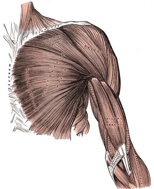
Every muscle in your body does exactly one thing: it shortens and pulls. It cannot push, cannot lengthen itself, and cannot choose a direction. Everything else — the precision of a surgeon's hand, the force of a sprint start, the fact that you can hold a cup without crushing it — comes from how those pulls are arranged, how finely they are graded, and how they are opposed by other muscles pulling the other way. Around 600 skeletal muscles make up roughly 40 per cent of body mass, and understanding them is mostly understanding leverage and control rather than strength.
The parts, and how they fit together
A skeletal muscle is a hierarchy of bundles inside bundles. The whole muscle is wrapped in epimysium; within it, fascicles are wrapped in perimysium; within each fascicle, individual muscle fibres are wrapped in endomysium. Those three connective tissue sheaths merge at the ends of the muscle to form the tendon, so force generated by any fibre anywhere in the belly is transmitted to bone. A muscle fibre is a single cell formed by the fusion of many myoblasts, which is why it carries dozens or hundreds of nuclei pressed against the inside of its membrane, and it can be several centimetres long.
Muscles attach at an origin, conventionally the more fixed end, and an insertion, the more mobile one, and they are named by a small vocabulary that once learned removes most of the memorising. Names describe shape (deltoid, trapezius, rhomboid), size (maximus, longus, brevis), fibre direction (rectus, oblique, transversus), location (temporalis, tibialis), number of heads (biceps, triceps, quadriceps) or action (flexor, extensor, adductor, levator). Fascicle arrangement determines the trade-off between range and force: parallel muscles such as sartorius shorten a long way but generate modest force, while pennate muscles such as deltoid pack many short fibres at an angle onto a tendon and generate much more force over a shorter range.
Muscles almost never act alone. The agonist, or prime mover, produces the intended movement; the antagonist opposes it and must relax, or contract in a controlled way, for the movement to be smooth; synergists assist and prevent unwanted movement at intervening joints; and fixators stabilise the origin so the pull is delivered where intended. Biceps and triceps at the elbow are the standard example, but the principle matters most where it is least obvious: the muscles stabilising your scapula do more work during a push-up than the ones visibly moving. Because a muscle can only pull, every joint needs opposing groups, and the resting tension of both is what we call muscle tone.
How the system works
Contraction is the sliding filament mechanism. Within each fibre, myofibrils are divided into repeating sarcomeres running from one Z line to the next, containing interdigitating thin actin and thick myosin filaments. A motor neuron's signal releases acetylcholine at the neuromuscular junction, depolarising the fibre; the impulse travels down T-tubules into the interior; calcium is released from the sarcoplasmic reticulum; calcium binds troponin, which shifts tropomyosin off the myosin binding sites on actin; myosin heads bind, pivot, and pull the thin filaments toward the centre, then detach using ATP and cycle again. The filaments themselves never shorten — they slide past one another. Relaxation is active too: calcium must be pumped back into the sarcoplasmic reticulum, which is why rigor mortis develops when ATP production stops and cross-bridges can neither detach nor release.
Force is graded in two ways, and this is what gives movement its precision. A motor unit is one motor neuron and all the fibres it supplies, and the ratio varies enormously: an extraocular muscle may have fewer than ten fibres per neuron, while gastrocnemius has over a thousand — which is exactly why the eye is precise and the calf is powerful. Recruiting more motor units increases force, and the smallest units are recruited first, so fine control is available at low force. Increasing firing frequency also increases force, because a second stimulus arriving before the first twitch has relaxed sums with it; at high enough frequency the twitches fuse into a smooth tetanic contraction. Skeletal muscle can do this because its action potential is far shorter than its twitch — unlike cardiac muscle, which cannot be tetanised at all.
Energy comes from three systems on different timescales, and the fibre types are built around them. A working muscle holds only a few seconds of ATP, backed by creatine phosphate for another eight to ten seconds — enough for a short sprint or a heavy lift. Anaerobic glycolysis then supplies energy for roughly the next minute or two, producing lactate. Beyond that, aerobic oxidation of glucose and fat in mitochondria supplies everything, and it is the only system that can run indefinitely. Type I fibres are slow, red with myoglobin, packed with mitochondria and capillaries, and fatigue-resistant — postural muscles are full of them. Type IIx fibres are fast, pale, glycolytic and fatigue quickly. Type IIa sit in between. Contractions are also classified by what happens to length: concentric when the muscle shortens, eccentric when it lengthens under load, which generates the most force and causes most delayed-onset soreness, and isometric when length does not change at all.
The cells that do the work
The contractile apparatus is built from a small set of proteins whose failures map directly onto disease. Actin and myosin do the work; titin, the largest protein in the body, runs from Z line to the centre of the sarcomere and acts as a molecular spring setting passive tension; troponin and tropomyosin form the calcium-operated switch. Dystrophin links the whole internal machinery through the cell membrane to the surrounding matrix, and its absence in Duchenne muscular dystrophy means every contraction tears the sarcolemma slightly. Skeletal muscle repairs itself using satellite cells, quiescent stem cells lying between the fibre membrane and its basal lamina, which activate after injury, proliferate, and fuse into the damaged fibre — adding nuclei rather than making new cells, since a fibre is already multinucleate. Training does not create new fibres in any meaningful number; hypertrophy is existing fibres adding myofibrils and nuclei. Fibre type proportions are largely inherited, which is a genuine part of why some people sprint and others run marathons, though endurance training does shift IIx fibres toward the more oxidative IIa profile.
Under the microscope
Skeletal muscle is one of the easiest tissues to identify and the three-question routine separates it from its relatives. In longitudinal section the fibres are long, straight, parallel cylinders that never branch, showing clear cross-striations from sarcomeres in register, with multiple flattened nuclei pressed against the periphery of each fibre immediately beneath the membrane. That peripheral, multinucleate arrangement is the decisive feature: cardiac muscle branches and has one central nucleus with dark intercalated discs, and smooth muscle has neither striations nor branching, just spindle-shaped cells with a single central nucleus. In transverse section skeletal muscle appears as a mosaic of polygonal fibres with nuclei around their edges, grouped into fascicles by perimysium, and this is the plane used clinically for muscle biopsy because it allows fibre size, shape and type to be assessed at once. Special stains for myosin ATPase separate type I from type II fibres and normally show a checkerboard mixture; loss of that checkerboard, with fibres of one type clustered together, indicates reinnervation after nerve damage.
When the system fails
Muscle disease and nerve disease produce different patterns, and telling them apart is the first clinical step. Myopathies typically cause symmetrical proximal weakness — difficulty rising from a chair, climbing stairs, or lifting arms overhead — with preserved reflexes and sensation, and a raised creatine kinase. Neuropathies more often cause distal weakness with sensory loss and reduced reflexes. Within the myopathies, Duchenne muscular dystrophy presents in early childhood with a waddling gait, calf pseudohypertrophy and grossly raised creatine kinase; inflammatory myopathies such as polymyositis and dermatomyositis present subacutely in adults; and statin-induced myopathy is common enough that muscle aching in a patient on a statin always deserves a creatine kinase check.
Acute breakdown of muscle is rhabdomyolysis, in which myoglobin from crushed or over-exerted fibres reaches the kidneys and precipitates in the tubules, causing acute kidney injury. The classic triad is muscle pain, weakness and dark tea-coloured urine, and the causes range from crush injury and prolonged immobility to extreme exertion, seizures, certain drugs and heat stroke. Compartment syndrome is a related surgical emergency: swelling within a fascial compartment raises pressure above capillary perfusion pressure, and the muscle inside it dies. Pain out of proportion to the injury and pain on passive stretch are the early signs, and waiting for absent pulses means waiting far too long.
Everyday muscle problems follow the same physiology. Delayed-onset muscle soreness peaks a day or two after unaccustomed exercise and follows eccentric contractions in particular, because lengthening under load produces the greatest force and the most microdamage. Cramp is sustained involuntary contraction, often related to fatigue or electrolyte disturbance. Disuse atrophy sets in within days of immobilisation and affects type I fibres first, which is why early mobilisation after surgery or critical illness matters so much more than it appears to. And because muscle is the body's largest reservoir of amino acids, prolonged catabolic illness breaks it down for fuel, producing the weakness that outlasts the illness itself.
Common questions about the muscular system
How do muscles work?
A motor neuron releases acetylcholine at the neuromuscular junction, the muscle fibre depolarises, and calcium is released inside it. Calcium moves tropomyosin off the binding sites on actin, allowing myosin heads to attach, pivot and pull the actin filaments toward the centre of each sarcomere, using ATP to detach and repeat. The filaments slide past each other rather than shortening. Relaxation requires calcium to be actively pumped back, which also costs ATP.
What are agonist and antagonist muscles?
The agonist, or prime mover, produces the intended movement; the antagonist lies on the opposite side of the joint and opposes it, relaxing or lengthening in a controlled way so the movement is smooth. Biceps and triceps at the elbow are the standard pair. Synergists assist and stop unwanted movement at other joints, and fixators hold the origin steady. Because muscle can only pull, opposing pairs are the only way to move a joint in both directions.
What is the difference between fast and slow muscle fibres?
Type I fibres are slow-twitch: red with myoglobin, rich in mitochondria and capillaries, energy from aerobic metabolism, and highly fatigue-resistant, so they dominate postural muscles and endurance activity. Type IIx fibres are fast-twitch: pale, glycolytic, powerful and quick to fatigue, used for sprinting and lifting. Type IIa are intermediate. The proportions are largely inherited, though endurance training shifts IIx fibres toward the more oxidative IIa profile.
Where do muscles get their energy?
From three systems on different timescales. Stored ATP lasts a few seconds and creatine phosphate extends that to roughly ten — enough for a short sprint or a heavy lift. Anaerobic glycolysis then covers the next minute or two and produces lactate. Beyond that, aerobic oxidation of glucose and fat in mitochondria supplies everything, and it is the only system that can continue indefinitely. Which system dominates is why sprinting and marathon running feel entirely different.
Selected references
- Hall JE, Hall ME. Guyton and Hall Textbook of Medical Physiology. 14th ed. Elsevier; 2021.
- Standring S, ed. Gray's Anatomy: The Anatomical Basis of Clinical Practice. 42nd ed. Elsevier; 2021.
- Ross MH, Pawlina W. Histology: A Text and Atlas. 8th ed. Wolters Kluwer; 2020.
- Schiaffino S, Reggiani C. Fiber types in mammalian skeletal muscles. Physiological Reviews. 2011;91(4):1447-1531.
- Dumont NA, Bentzinger CF, Sincennes MC, Rudnicki MA. Satellite cells and skeletal muscle regeneration. Comprehensive Physiology. 2015;5(3):1027-1059.
- Kumar V, Abbas AK, Aster JC. Robbins and Cotran Pathologic Basis of Disease. 10th ed. Elsevier; 2021.
Medical disclaimer. This article is written for education and general understanding. It is not medical advice and cannot replace assessment by a qualified healthcare professional. If you have symptoms or health concerns, speak with a clinician. Read the full disclaimer.