Breathing looks like the simplest thing the body does and is one of the most finely engineered. Air has to be warmed, humidified and filtered, delivered through a branching tree that divides about twenty-three times, and then handed across a barrier so thin that it is measured in fractions of a micrometre — all without the lung collapsing under its own surface tension, and all regulated so precisely that arterial carbon dioxide varies by only a few per cent between sleep and a sprint. The system is best understood in two halves: the plumbing that moves air, and the membrane where the actual work happens.
The parts, and how they fit together
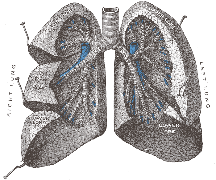
The conducting zone moves air and conditions it but exchanges no gas. It begins at the nose, where turbinates create turbulence that warms and humidifies incoming air and traps particles, continues through the pharynx and larynx, and reaches the trachea, which is held open by around twenty C-shaped cartilage rings with a muscular posterior wall that lets the oesophagus bulge into it when you swallow. The trachea divides into two main bronchi at the carina, and the right one is wider, shorter and more vertical, which is why inhaled objects lodge on the right far more often than the left. Bronchi divide into bronchioles, which lose their cartilage and depend on the surrounding lung tissue to stay open, and end as terminal bronchioles.
The respiratory zone is where gas exchange happens and begins at the respiratory bronchioles. These lead into alveolar ducts and finally alveolar sacs, and the numbers are the striking part: roughly 300 to 500 million alveoli give a total surface area of around 70 square metres, close to half a tennis court, folded into a space the size of your chest. Each alveolus is wrapped in a capillary net so dense that blood flows over it almost as a continuous sheet, and the barrier between air and blood is only about 0.2 to 0.6 micrometres thick — type I pneumocyte, fused basement membrane, capillary endothelium.
The mechanics belong to the chest wall, not the lungs. The lungs have no skeletal muscle of their own and are moved passively. The diaphragm, a dome of muscle supplied by the phrenic nerves from cervical roots three, four and five, does about 70 per cent of quiet breathing by descending and increasing the vertical dimension of the thorax. The external intercostals lift the ribs. Each lung sits in a pleural cavity lined by visceral and parietal pleura separated by a thin film of fluid, and the negative pressure in that space is what keeps the lung expanded against its own elastic recoil — which is why puncturing it causes the lung to collapse.
How the system works
Ventilation is driven entirely by pressure differences. When the diaphragm contracts, thoracic volume rises, intrapleural pressure becomes more negative, alveolar pressure falls just below atmospheric, and air flows in. Quiet expiration requires no muscular effort at all: the stretched elastic tissue of the lung recoils and pushes the air out, which is why expiration is normally longer and more passive than inspiration. Forced expiration recruits the abdominal muscles and internal intercostals. Two forces oppose inflation — the elastic recoil of the tissue, and the surface tension of the fluid lining each alveolus, which is by far the larger. Type II pneumocytes solve the second by secreting surfactant, a phospholipid that lowers surface tension and, crucially, lowers it more in small alveoli than large ones, which stops small alveoli emptying into big ones.
Gas exchange itself is passive diffusion down partial pressure gradients. Alveolar air holds oxygen at about 100 millimetres of mercury against 40 in the arriving venous blood, so oxygen crosses; carbon dioxide sits at 45 in the blood against 40 in the alveolus, a much smaller gradient, but carbon dioxide is around twenty times more soluble so it diffuses just as effectively. Blood spends about 0.75 seconds in a pulmonary capillary at rest and equilibrates in roughly a third of that, which is why healthy lungs have a large reserve and why oxygen saturation only falls once disease is well advanced. Matching matters as much as diffusion: regions that are ventilated but not perfused, or perfused but not ventilated, waste effort, and the lung actively constricts arterioles in poorly ventilated regions to redirect blood — hypoxic pulmonary vasoconstriction, the opposite of what every other organ does.
Once in the blood, about 98.5 per cent of oxygen binds haemoglobin and the rest dissolves in plasma. Haemoglobin's four subunits bind cooperatively, producing the sigmoid dissociation curve that lets blood load almost fully in the lung and unload readily in tissue; the curve shifts right in warm, acidic, carbon-dioxide-rich conditions, so exercising muscle automatically extracts more. Carbon dioxide returns mostly as bicarbonate generated inside red cells by carbonic anhydrase. Control of breathing sits in the medulla and pons, and the primary stimulus is not oxygen but carbon dioxide: central chemoreceptors respond to the pH of cerebrospinal fluid, which tracks arterial carbon dioxide closely. Peripheral chemoreceptors in the carotid and aortic bodies do respond to oxygen, but only strongly once arterial oxygen falls below about 60 millimetres of mercury, which is why holding your breath becomes unbearable from carbon dioxide long before you are short of oxygen.
The cells that do the work
Three cell types define the exchange surface, and a fourth defines the airway. Type I pneumocytes are extraordinarily flattened squamous cells that cover around 95 per cent of the alveolar surface while making up only about 40 per cent of the cells, and their thinness is the whole point — they are the air side of the barrier. Type II pneumocytes are cuboidal, far more numerous, and do two jobs: they synthesise and secrete surfactant from lamellar bodies, and they act as the stem cell of the alveolus, dividing to replace type I cells after injury. Alveolar macrophages, sometimes called dust cells, patrol the alveolar surface and are the last line of defence for anything that got past the mucociliary escalator. That escalator is the airway's own mechanism: pseudostratified ciliated columnar epithelium with goblet cells produces a mucus layer that traps particles, and several hundred cilia on each cell beat in coordinated waves at ten to twenty hertz, moving the sheet upward toward the pharynx to be swallowed. Cigarette smoke paralyses those cilia within minutes and eventually replaces the epithelium with a stratified squamous one that has none, which is why a smoker's cough is productive and worse in the morning.
Under the microscope
Moving down the airway on a slide, the changes come in a strict and predictable sequence, and knowing it lets you place any section. The trachea and bronchi are lined by pseudostratified ciliated columnar epithelium with goblet cells, supported by cartilage — rings in the trachea, irregular plates in the bronchi — with seromucous glands in the submucosa and a band of smooth muscle. Bronchioles are defined by what they have lost: no cartilage, no glands, and progressively fewer goblet cells, with a proportionally thicker smooth muscle layer, which is precisely why they are the airways that constrict in asthma. Epithelium thins from simple columnar to simple cuboidal and gains club cells, which secrete protective proteins and act as a local stem cell. Respiratory bronchioles are recognised by alveoli budding directly off their walls — the transition point. Alveolar walls are almost nothing: a sheet of type I pneumocytes with occasional plump type II cells bulging at the corners, a shared basement membrane, capillary endothelium, and a scaffold of elastic and reticular fibres. Alveolar pores of Kohn connect adjacent alveoli and allow collateral ventilation past a blocked bronchiole.
When the system fails
Obstructive diseases limit airflow out. Chronic obstructive pulmonary disease combines emphysema, in which alveolar walls are destroyed and elastic recoil is lost so the airways collapse on expiration, with chronic bronchitis, in which inflammation and mucus narrow the lumen; the result is air trapping, a prolonged expiratory phase and a reduced FEV1 to FVC ratio. Asthma is also obstructive but is characterised by reversible bronchoconstriction, airway inflammation and hyperresponsiveness rather than permanent destruction, which is why bronchodilators help so much more in asthma than in emphysema. Restrictive diseases do the opposite, limiting how much the lung can expand, whether through fibrosis of the tissue itself or through chest wall and neuromuscular problems.
Failures of the exchange surface present as hypoxia. Pneumonia fills alveoli with inflammatory exudate so that blood passes through unventilated tissue — a shunt, which is why oxygen therapy alone helps less than expected. Pulmonary oedema does the same with fluid, usually because left heart failure has raised pulmonary capillary pressure. Acute respiratory distress syndrome is diffuse alveolar damage from sepsis, trauma or aspiration, in which the barrier leaks protein-rich fluid and hyaline membranes form. Pulmonary embolism is the opposite mismatch: alveoli are ventilated but not perfused, producing sudden breathlessness and chest pain with a chest X-ray that can look almost normal.
Two mechanical failures are worth recognising immediately. A pneumothorax is air in the pleural space, which abolishes the negative pressure holding the lung expanded and lets it collapse; a tension pneumothorax, in which air enters but cannot escape, progressively compresses the mediastinum and is an emergency treated by decompression before imaging. Because the phrenic nerves arise from cervical roots three to five, a spinal cord injury above that level paralyses the diaphragm and stops spontaneous breathing entirely, while an injury below it may leave breathing intact — the origin of the clinical aphorism that C3, 4 and 5 keep the diaphragm alive.
Common questions about the respiratory system
How does the respiratory system work?
The diaphragm and intercostal muscles enlarge the chest, which lowers pressure inside the lungs and draws air in through a branching airway that warms, humidifies and filters it. At the alveoli, oxygen diffuses into blood and carbon dioxide diffuses out, down their partial pressure gradients. Quiet expiration needs no muscular effort at all — the stretched elastic lung simply recoils. The rate is set in the brainstem, primarily by arterial carbon dioxide.
What is the difference between the conducting and respiratory zones?
The conducting zone — nose, pharynx, larynx, trachea, bronchi and bronchioles down to the terminal bronchioles — moves air and conditions it but exchanges no gas, so it is anatomical dead space, about 150 millilitres in an adult. The respiratory zone begins at the respiratory bronchioles, where alveoli start budding off the walls, and includes the alveolar ducts and sacs. That is where all gas exchange happens.
Why do we need surfactant?
Because surface tension in the fluid lining each alveolus is a far bigger obstacle to inflation than the elasticity of the tissue, and because without it small alveoli would empty into large ones. Surfactant, a phospholipid secreted by type II pneumocytes, lowers surface tension and lowers it more in small alveoli than large ones, which keeps them all open at similar sizes. Premature babies born before surfactant production is established develop respiratory distress syndrome for exactly this reason.
Why do inhaled objects usually go into the right lung?
Because the right main bronchus is wider, shorter and more vertical than the left, which angles away to make room for the heart. An object falling past the carina therefore follows the straighter path. The same anatomy explains why aspiration pneumonia most often affects the right lower lobe in someone lying on their back.
Selected references
- West JB, Luks AM. West's Respiratory Physiology: The Essentials. 11th ed. Wolters Kluwer; 2021.
- Hall JE, Hall ME. Guyton and Hall Textbook of Medical Physiology. 14th ed. Elsevier; 2021.
- Standring S, ed. Gray's Anatomy: The Anatomical Basis of Clinical Practice. 42nd ed. Elsevier; 2021.
- Ross MH, Pawlina W. Histology: A Text and Atlas. 8th ed. Wolters Kluwer; 2020.
- Whitsett JA, Wert SE, Weaver TE. Diseases of pulmonary surfactant homeostasis. Annual Review of Pathology. 2015;10:371-393.
- Kumar V, Abbas AK, Aster JC. Robbins and Cotran Pathologic Basis of Disease. 10th ed. Elsevier; 2021.
Medical disclaimer. This article is written for education and general understanding. It is not medical advice and cannot replace assessment by a qualified healthcare professional. If you have symptoms or health concerns, speak with a clinician. Read the full disclaimer.