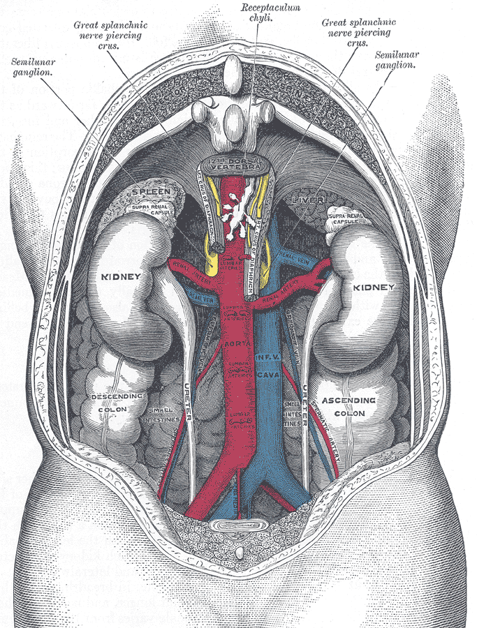
The kidneys are the body's regulators of internal chemistry. Together they weigh about 300 grams, roughly 0.4 percent of body mass, yet they receive around 20 percent of cardiac output because their function depends entirely on continuous perfusion. Each day they filter roughly 180 litres of plasma and return more than 99 percent of it, adjusting water, sodium, potassium, calcium, phosphate, and acid-base balance minute by minute so that the composition of the internal environment stays almost constant.
Anatomy: the structure
The kidneys are retroperitoneal, lying against the posterior abdominal wall between roughly the T12 and L3 vertebral levels, with the right kidney sitting slightly lower because of the liver. Each is wrapped in a fibrous capsule, then perirenal fat, renal fascia, and pararenal fat, which together cushion and anchor it. At the hilum, structures are arranged from anterior to posterior as vein, artery, and ureter.
A coronal section shows an outer cortex and an inner medulla containing 8 to 18 renal pyramids. Each pyramid drains through a papilla into a minor calyx; minor calyces merge into major calyces, which merge into the renal pelvis and then the ureter. Cortical tissue extends between pyramids as the renal columns of Bertin, carrying interlobar vessels.
Each kidney contains about one million nephrons, and each nephron begins with a renal corpuscle made of a glomerular capillary tuft inside Bowman capsule. Filtrate then passes through the proximal convoluted tubule, the descending and ascending limbs of the loop of Henle, the distal convoluted tubule, and the collecting duct. Cortical nephrons have short loops, while the roughly fifteen percent that are juxtamedullary have long loops descending deep into the medulla and are responsible for producing concentrated urine. The vasculature is unusual in having two capillary beds in series: the glomerulus between afferent and efferent arterioles, then the peritubular capillaries and vasa recta.
Physiology: how it works
Filtration is driven by Starling forces across the glomerular barrier. Glomerular capillary hydrostatic pressure of roughly 55 mmHg is opposed by capsular hydrostatic pressure of about 15 mmHg and plasma oncotic pressure of about 30 mmHg, leaving a net filtration pressure near 10 mmHg and a glomerular filtration rate of about 125 mL/min. The barrier has three layers, fenestrated endothelium, a fused basement membrane rich in negatively charged heparan sulfate, and podocyte filtration slits, so filtration is selective by both size and charge, which is why albumin is normally retained.
Reabsorption is segmental and specific. The proximal convoluted tubule reclaims about 65 percent of filtered sodium and water plus essentially all glucose and amino acids, driven by the basolateral sodium-potassium ATPase. The thin descending limb is permeable to water but not solute, the thick ascending limb is the reverse and actively pumps sodium, potassium, and chloride through the NKCC2 transporter, and this separation of permeabilities creates the countercurrent multiplier that establishes a medullary osmotic gradient rising from 300 to about 1200 mOsm/kg.
The final adjustment happens distally under hormonal control. Antidiuretic hormone inserts aquaporin-2 channels into the collecting duct, allowing water to follow the medullary gradient and concentrate the urine. Aldosterone increases sodium reabsorption and potassium secretion in the principal cells. The juxtaglomerular apparatus, where the macula densa of the distal tubule contacts its own glomerulus, senses distal sodium delivery and releases renin, initiating the renin-angiotensin-aldosterone cascade. The kidneys also secrete erythropoietin in response to hypoxia and perform the final hydroxylation that activates vitamin D.
Biology: the living cells
Renal cell biology is a study in specialisation by segment. Podocytes wrap the glomerular capillary with interdigitating foot processes bridged by nephrin-based slit diaphragms, and effacement of those foot processes is the structural signature of nephrotic proteinuria. Proximal tubule cells carry a dense brush border of microvilli that multiplies surface area, abundant mitochondria for active transport, and endocytic machinery for reclaiming filtered protein. Thick ascending limb cells are mitochondria-rich but lack water channels, which is exactly what the countercurrent mechanism requires. In the collecting duct, principal cells handle sodium, potassium, and water under aldosterone and antidiuretic hormone control, while intercalated cells manage acid-base balance, with type A cells secreting protons and type B cells secreting bicarbonate. Juxtaglomerular cells are modified smooth muscle cells in the afferent arteriole that store and release renin.
Histology: under the microscope
A cortical section shows rounded renal corpuscles surrounded by densely packed tubules. Proximal tubules are recognised by their brush border, deeply eosinophilic cytoplasm, and indistinct lumen; distal tubules have a clearer lumen, paler cytoplasm, no brush border, and more visible nuclei. Within the corpuscle, the vascular pole carries the afferent and efferent arterioles and the urinary pole opens into the proximal tubule, and mesangial cells provide structural support and phagocytic capacity between capillary loops. The medulla is dominated by parallel straight structures: thin and thick limbs of Henle, collecting ducts, and the vasa recta, all running toward the papilla. Collecting ducts are identified by their distinct cell borders and pale cuboidal to columnar epithelium, and at the papillary tip the epithelium transitions to the urothelium of the calyx.
Clinical correlations
Acute kidney injury is classified by where the problem lies. Prerenal injury reflects reduced perfusion from hypovolaemia or low cardiac output and is reversible if perfusion is restored quickly. Intrinsic injury damages the parenchyma itself, most commonly as acute tubular necrosis affecting the vulnerable proximal tubule and thick ascending limb. Postrenal injury results from obstruction anywhere from the renal pelvis to the urethra, and relieving it early prevents permanent damage.
Glomerular disease presents in two broad patterns. Nephrotic syndrome involves podocyte injury with heavy proteinuria above 3.5 grams per day, hypoalbuminaemia, oedema, and hyperlipidaemia. Nephritic syndrome involves inflammation of the glomerulus with haematuria, red cell casts, hypertension, and a falling filtration rate. The distinction is structural: nephrotic disease damages the filtration barrier, nephritic disease inflames and disrupts the capillary wall.
Chronic kidney disease is staged by estimated glomerular filtration rate and albuminuria, and its complications follow directly from lost renal functions. Reduced erythropoietin produces anaemia, impaired vitamin D activation and phosphate retention produce mineral and bone disorder with secondary hyperparathyroidism, impaired acid excretion produces metabolic acidosis, and impaired sodium and water handling produces hypertension and volume overload. Diuretics act on identifiable transporters, with loop diuretics blocking NKCC2 in the thick ascending limb and thiazides blocking the sodium-chloride cotransporter in the distal convoluted tubule.
Common questions about the kidneys
What is a nephron and what does it do?
The nephron is the functional unit of the kidney, and each kidney holds roughly a million of them. It begins with a renal corpuscle that filters plasma, then a tubular system that selectively reabsorbs water and useful solutes back into blood and secretes additional waste, producing urine as the final adjusted output.
How do the kidneys concentrate urine?
The loop of Henle acts as a countercurrent multiplier: the descending limb is permeable to water while the ascending limb actively pumps out salt without water following. This builds an osmotic gradient in the medulla, and antidiuretic hormone then makes the collecting duct permeable so water leaves the filtrate as it passes through that gradient.
Why is protein in the urine a warning sign?
The glomerular filtration barrier normally excludes albumin by both size and negative charge. Persistent proteinuria indicates that this barrier is damaged, most often through injury to podocytes or to the basement membrane, and it is one of the earliest and strongest predictors of progressive kidney disease.
What hormones do the kidneys produce?
The kidneys release renin from juxtaglomerular cells to regulate blood pressure through the renin-angiotensin-aldosterone system, erythropoietin in response to low oxygen to stimulate red cell production, and the enzyme that performs the final activation step of vitamin D, which controls calcium absorption.
Selected references
- Eaton DC, Pooler JP. Vander's Renal Physiology. 9th ed. McGraw Hill; 2018.
- Hall JE, Hall ME. Guyton and Hall Textbook of Medical Physiology. 14th ed. Elsevier; 2021.
- Standring S, ed. Gray's Anatomy: The Anatomical Basis of Clinical Practice. 42nd ed. Elsevier; 2021.
- Ross MH, Pawlina W. Histology: A Text and Atlas. 8th ed. Wolters Kluwer; 2020.
- Boron WF, Boulpaep EL. Medical Physiology. 3rd ed. Elsevier; 2017.
- Kidney Disease: Improving Global Outcomes (KDIGO) CKD Work Group. KDIGO 2024 Clinical Practice Guideline for the Evaluation and Management of Chronic Kidney Disease. Kidney International. 2024.
Medical disclaimer. This article is written for education and general understanding. It is not medical advice and cannot replace assessment by a qualified healthcare professional. If you have symptoms or health concerns, speak with a clinician. Read the full disclaimer.