Breathing looks simple from the outside, but each breath is a precisely engineered exchange between the atmosphere and the bloodstream. The lungs deliver oxygen to roughly 300 million alveoli, remove carbon dioxide, and act as one of the body's two main systems for regulating blood pH. Their branching design packs a gas-exchange surface of about 70 square metres into a space small enough to fit inside the rib cage.
Anatomy: the structure
Air travels from the nose and pharynx through the larynx into the trachea, which divides at the carina into right and left main bronchi. The right main bronchus is wider, shorter, and more vertical, which is why inhaled foreign bodies enter it more often. Each main bronchus divides into lobar then segmental bronchi, and division continues for roughly 23 generations, ending in the alveolar sacs. Cartilage supports the larger airways, gradually disappears from the bronchioles, and is replaced by smooth muscle that controls airway calibre.
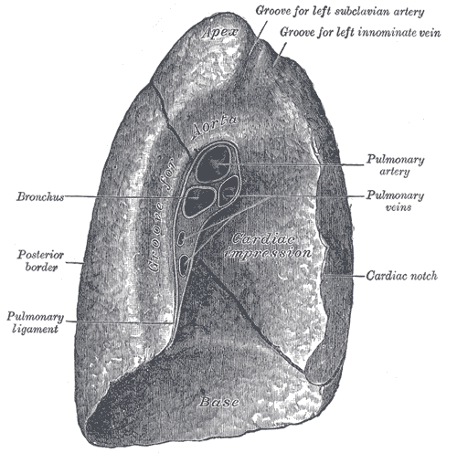
The right lung has three lobes separated by the oblique and horizontal fissures; the left lung has two lobes and a cardiac notch that accommodates the heart. Each lung is enclosed by a double-layered pleura: the visceral pleura adheres to the lung surface, the parietal pleura lines the chest wall and diaphragm, and a thin layer of pleural fluid between them couples the lung to the chest wall while allowing frictionless sliding.
The lungs receive two separate blood supplies. The pulmonary arteries carry deoxygenated blood from the right ventricle to the alveolar capillary bed at low pressure, roughly one-sixth of systemic pressure. The bronchial arteries, arising from the aorta, carry oxygenated blood at systemic pressure to nourish the airway walls themselves. Beyond the terminal bronchioles lies the respiratory zone: respiratory bronchioles, alveolar ducts, and alveolar sacs, the only regions where gas exchange actually occurs.
Physiology: how it works
Inspiration is active. The diaphragm contracts and flattens, the external intercostals lift the ribs, thoracic volume increases, and intrapleural pressure becomes more negative, so alveolar pressure falls below atmospheric pressure and air flows inward. Quiet expiration is passive, driven by the elastic recoil of lung tissue and the surface tension of the alveolar lining; forced expiration recruits the abdominal wall and internal intercostals.
Gas exchange is pure diffusion down partial pressure gradients across a respiratory membrane less than a micrometre thick. Alveolar oxygen at about 104 mmHg meets capillary blood at about 40 mmHg, and carbon dioxide moves the other way across a much smaller gradient, which its higher solubility more than compensates for. Efficiency depends on matching ventilation to perfusion, and hypoxic pulmonary vasoconstriction helps by diverting blood away from poorly ventilated regions.
Oxygen is carried almost entirely by haemoglobin, and the sigmoid oxygen-haemoglobin dissociation curve means saturation stays high across a wide range of alveolar pressures while unloading readily in active tissue. Carbon dioxide travels mostly as bicarbonate generated by carbonic anhydrase inside red cells. Because that reaction is reversible, the rate of ventilation directly sets arterial carbon dioxide and therefore blood pH, and central chemoreceptors in the medulla respond primarily to that signal.
Biology: the living cells
The alveolar surface is built from two cell types with very different jobs. Type I pneumocytes are extremely flattened squamous cells that cover about 95 percent of the alveolar surface area and create a diffusion barrier thin enough for gas to cross almost instantly. Type II pneumocytes are cuboidal, far more numerous by count, and synthesise pulmonary surfactant, a phospholipid and protein mixture that lowers alveolar surface tension, prevents small alveoli from collapsing into larger ones, and dramatically reduces the work of breathing. Type II cells also serve as the progenitor population that regenerates the alveolar lining after injury. Alveolar macrophages patrol the air spaces, clearing inhaled particles and pathogens, while the conducting airways are protected by the mucociliary escalator formed by ciliated cells and goblet cells.
Histology: under the microscope
Moving down the airway, histology changes in a predictable sequence. The trachea and bronchi are lined by pseudostratified ciliated columnar epithelium with goblet cells, supported by cartilage rings or plates and seromucous glands. Bronchioles lose their cartilage and goblet cells, shift to simple columnar then simple cuboidal epithelium, and gain club cells that secrete protective proteins. In the respiratory zone the wall thins to almost nothing: alveolar septa contain type I and type II pneumocytes, a fused basement membrane, capillary endothelium, and a scaffold of elastic and reticular fibres that stores the energy released during expiration. Small openings called alveolar pores allow collateral ventilation between adjacent alveoli.
Clinical correlations
Chronic obstructive pulmonary disease combines emphysema, in which alveolar walls are destroyed and elastic recoil is lost, with chronic bronchitis, in which airway inflammation and mucus hypersecretion narrow the lumen. The result is air trapping, a prolonged expiratory phase, and a reduced FEV1/FVC ratio. Asthma is also obstructive but is characterised by reversible bronchoconstriction, airway inflammation, and hyperresponsiveness rather than permanent tissue destruction.
Restrictive disease works the opposite way. In pulmonary fibrosis, excess collagen deposition stiffens the interstitium, reduces compliance, and thickens the respiratory membrane, so lung volumes fall and diffusion capacity drops. Respiratory distress syndrome of the newborn is a surfactant deficiency: without adequate surfactant, alveolar surface tension is unopposed, alveoli collapse at end-expiration, and the work of breathing rises sharply.
Pneumonia fills alveoli with inflammatory exudate, creating a ventilation-perfusion mismatch and a physiological shunt. Pulmonary embolism does the reverse, blocking perfusion to ventilated lung and increasing dead space. Pneumothorax breaks the pleural seal, allowing air into the pleural space so the lung recoils inward and the chest wall springs outward, which is why the negative intrapleural pressure is essential rather than incidental.
Common questions about the lungs
How does oxygen get from the air into the blood?
Oxygen diffuses down its partial pressure gradient from alveolar air across the respiratory membrane into pulmonary capillary blood. The membrane is only about half a micrometre thick, so equilibration takes roughly a quarter of a second, and haemoglobin inside red blood cells then binds the oxygen for transport.
What does surfactant do and why does it matter?
Surfactant is a phospholipid and protein mixture made by type II alveolar cells. It lowers the surface tension of the fluid lining the alveoli, which prevents small alveoli from collapsing into larger ones and substantially reduces the muscular work needed to inflate the lungs with each breath.
Why does the right lung have three lobes and the left only two?
The heart sits slightly left of the midline and occupies space in the left hemithorax, producing the cardiac notch. The left lung is correspondingly smaller and divided by a single oblique fissure into two lobes, while the right lung is divided by oblique and horizontal fissures into three.
What is the difference between the conducting zone and the respiratory zone?
The conducting zone runs from the nose to the terminal bronchioles and warms, humidifies, and filters air without exchanging gas, forming the anatomical dead space. The respiratory zone begins at the respiratory bronchioles and includes alveolar ducts and sacs, where gas exchange actually takes place.
Selected references
- West JB, Luks AM. West's Respiratory Physiology: The Essentials. 11th ed. Wolters Kluwer; 2021.
- Standring S, ed. Gray's Anatomy: The Anatomical Basis of Clinical Practice. 42nd ed. Elsevier; 2021.
- Hall JE, Hall ME. Guyton and Hall Textbook of Medical Physiology. 14th ed. Elsevier; 2021.
- Ross MH, Pawlina W. Histology: A Text and Atlas. 8th ed. Wolters Kluwer; 2020.
- Weibel ER. What makes a good lung? Swiss Medical Weekly. 2009;139(27-28):375-386.
- Mescher AL. Junqueira's Basic Histology: Text and Atlas. 16th ed. McGraw Hill; 2021.
Medical disclaimer. This article is written for education and general understanding. It is not medical advice and cannot replace assessment by a qualified healthcare professional. If you have symptoms or health concerns, speak with a clinician. Read the full disclaimer.