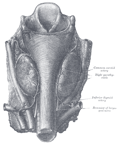
The thyroid is a small gland with an unusually wide reach. Weighing only 15 to 25 grams, it produces hormones that set the basal metabolic rate of nearly every cell, influence growth, and are indispensable for normal brain development in the first years of life. It is also the only endocrine gland that stores its product extracellularly, holding enough hormone in reserve to last several months.
Anatomy: the structure
The thyroid sits in the anterior neck at the level of the C5 to T1 vertebrae, with right and left lobes joined across the midline by an isthmus lying over the second to fourth tracheal rings. A pyramidal lobe extending upward from the isthmus is present in roughly half of people, a remnant of the thyroglossal duct along which the gland descended from the foramen caecum of the tongue during development. Because the gland is enclosed in the pretracheal fascia and attached to the larynx, it moves upward on swallowing, which is the classic clinical test for identifying a thyroid mass.
Its relations explain the risks of thyroid surgery. The recurrent laryngeal nerves ascend in the tracheo-oesophageal groove close to the inferior thyroid artery and supply all intrinsic laryngeal muscles except the cricothyroid; injury causes hoarseness, or airway obstruction if bilateral. The external branch of the superior laryngeal nerve runs near the superior thyroid artery and supplies cricothyroid, so injury alters voice pitch. The four parathyroid glands lie on the posterior surface of the lobes and can be removed inadvertently, causing hypocalcaemia.
The gland is one of the most richly perfused tissues in the body relative to its mass. The superior thyroid artery, the first branch of the external carotid, supplies the upper pole, and the inferior thyroid artery from the thyrocervical trunk supplies the lower pole. Superior and middle thyroid veins drain into the internal jugular vein while the inferior thyroid veins drain into the brachiocephalic veins. Functionally, the gland is built from roughly one million spherical follicles, each a single layer of epithelium enclosing a lumen filled with colloid.
Physiology: how it works
Hormone synthesis is a multi-step process built around iodine. Follicular cells actively concentrate iodide from blood using the sodium-iodide symporter, achieving intracellular levels twenty to forty times higher than plasma. At the apical membrane, thyroid peroxidase oxidises iodide and attaches it to tyrosine residues on thyroglobulin, forming monoiodotyrosine and diiodotyrosine. The same enzyme then couples these to form T4 from two diiodotyrosines or T3 from one of each, and the product is stored within thyroglobulin in the colloid until needed.
Release reverses the process. Under TSH stimulation, follicular cells endocytose colloid, lysosomal proteases digest thyroglobulin, and free T4 and T3 are released into the circulation in roughly a twenty to one ratio. T4 is essentially a prohormone with a half-life of about seven days; peripheral deiodinase enzymes in liver, kidney, and target tissues convert it to the three to four times more potent T3, or to inactive reverse T3 when metabolic demand is low. Over 99 percent of circulating hormone is bound to thyroxine-binding globulin, transthyretin, and albumin, so only the free fraction is active.
Control is a classic negative feedback loop. The hypothalamus releases TRH, the anterior pituitary responds with TSH, and TSH stimulates every step from iodide uptake to hormone release as well as the growth of the gland itself. Circulating free T3 and T4 inhibit both TRH and TSH release, which is why TSH is the most sensitive single measure of thyroid status and moves in the opposite direction to the hormone level. At the tissue level, T3 enters the nucleus, binds thyroid hormone receptors on DNA, and alters transcription, increasing sodium-potassium ATPase activity and mitochondrial oxidative capacity, raising basal metabolic rate and heat production, and increasing the density of beta-adrenergic receptors so that tissues respond more strongly to catecholamines.
Biology: the living cells
Two endocrine cell types share the gland. Follicular cells are polarised epithelial cells with a basal membrane carrying the sodium-iodide symporter and the TSH receptor, and an apical membrane carrying thyroid peroxidase and pendrin. They are unusual in performing both synthesis and, later, retrieval and digestion of their own secretory product, so they carry abundant rough endoplasmic reticulum and Golgi for thyroglobulin export and abundant lysosomes for its later breakdown. Their height changes with activity: flattened and inactive when TSH is low, tall columnar with scalloped colloid margins when strongly stimulated. Parafollicular or C cells are larger, paler, neural crest derived, and lie between follicles or within the follicular basement membrane without contacting the colloid. They secrete calcitonin in response to high plasma calcium, which inhibits osteoclasts, although in adult humans its physiological role is minor compared with parathyroid hormone. C cells are clinically important because they give rise to medullary thyroid carcinoma.
Histology: under the microscope
Thyroid tissue is immediately recognisable as a honeycomb of closed spherical follicles, a pattern seen in no other gland, because the secretory product is stored outside the cells rather than within them. Each follicle is lined by a single layer of epithelium, typically simple cuboidal in the resting state, surrounding a lumen of homogeneous eosinophilic colloid consisting mainly of thyroglobulin. Follicle size and epithelial height are a direct readout of activity: a hypoactive gland shows large follicles distended with colloid and flattened epithelium, while a hyperactive gland shows small follicles, reduced colloid, tall columnar cells, and scalloped resorption lacunae at the colloid margin where endocytosis is under way. Parafollicular C cells appear as clear cells in the interfollicular spaces and are best confirmed with calcitonin immunostaining. A dense capillary network surrounds every follicle, and connective tissue septa divide the gland into lobules.
Clinical correlations
Hypothyroidism slows almost everything. Fatigue, cold intolerance, weight gain, constipation, bradycardia, dry skin, and slowed reflex relaxation follow from reduced metabolic rate and reduced adrenergic sensitivity. Hashimoto thyroiditis, an autoimmune lymphocytic infiltration with antibodies to thyroid peroxidase, is the commonest cause where iodine intake is adequate; worldwide, dietary iodine deficiency remains the leading cause, and untreated congenital hypothyroidism causes irreversible intellectual disability, which is why newborn screening is routine.
Hyperthyroidism does the reverse. Weight loss despite good appetite, heat intolerance, tremor, anxiety, palpitations, and atrial fibrillation reflect a raised metabolic rate and increased beta-adrenergic sensitivity, which is why beta blockers relieve symptoms rapidly without altering hormone levels. Graves disease is the commonest cause, driven by stimulating antibodies against the TSH receptor, and it produces diffuse goitre with the additional signs of ophthalmopathy and pretibial myxoedema that toxic nodular disease does not.
Interpreting thyroid function starts with TSH because of the feedback loop: a raised TSH with low free T4 indicates primary hypothyroidism, while a suppressed TSH with raised free T4 indicates primary hyperthyroidism, and discordant patterns point to pituitary or hypothalamic disease. Thyroid nodules are common and mostly benign, assessed by ultrasound and fine needle aspiration; papillary carcinoma is the most frequent malignancy and has characteristic nuclear features and a generally good prognosis, while medullary carcinoma arising from C cells is monitored using calcitonin.
Common questions about the thyroid
What is the difference between T3 and T4?
T4 is the main product of the gland and acts largely as a circulating prohormone with a long half-life. T3 is three to four times more potent and is mostly produced outside the thyroid when deiodinase enzymes in peripheral tissues remove one iodine atom from T4, which lets each tissue adjust its own active hormone supply.
Why is TSH the first test for thyroid problems?
The pituitary responds very sensitively to small changes in circulating free thyroid hormone, so TSH moves before free T4 leaves the normal range. It also moves in the opposite direction: a high TSH indicates an underactive gland and a suppressed TSH indicates an overactive one, provided the pituitary itself is healthy.
Why does the thyroid need iodine?
Iodine is a structural component of the hormones themselves; T4 contains four iodine atoms and T3 contains three. Without adequate dietary iodine the gland cannot synthesise hormone, TSH rises in an attempt to compensate, and the sustained stimulation enlarges the gland into a goitre.
What does an overactive thyroid look like under the microscope?
Follicles become smaller, the epithelium becomes tall and columnar rather than cuboidal, colloid is reduced, and scalloped resorption lacunae appear at the edge of the colloid where the cells are actively endocytosing it. An underactive gland shows the opposite: large follicles packed with colloid and flattened epithelium.
Selected references
- Standring S, ed. Gray's Anatomy: The Anatomical Basis of Clinical Practice. 42nd ed. Elsevier; 2021.
- Melmed S, et al. Williams Textbook of Endocrinology. 14th ed. Elsevier; 2019.
- Hall JE, Hall ME. Guyton and Hall Textbook of Medical Physiology. 14th ed. Elsevier; 2021.
- Ross MH, Pawlina W. Histology: A Text and Atlas. 8th ed. Wolters Kluwer; 2020.
- Bianco AC, et al. Deiodinases: implications of the local control of thyroid hormone action. Journal of Clinical Investigation. 2006;116(10):2571-2579.
- Kumar V, Abbas AK, Aster JC. Robbins and Cotran Pathologic Basis of Disease. 10th ed. Elsevier; 2021.
Medical disclaimer. This article is written for education and general understanding. It is not medical advice and cannot replace assessment by a qualified healthcare professional. If you have symptoms or health concerns, speak with a clinician. Read the full disclaimer.