Calling this the waste disposal system undersells it considerably. Removing urea is the least interesting thing the kidneys do. They set the volume and composition of every fluid compartment in the body, control blood pressure over the long term, determine blood pH, decide how many red cells you make, and perform the final activation of vitamin D. Urine is simply what is left after all that regulation has happened — the residue of a set of decisions rather than the purpose of the organ. The rest of the system exists to store that residue and get rid of it at a socially convenient moment.
The parts, and how they fit together
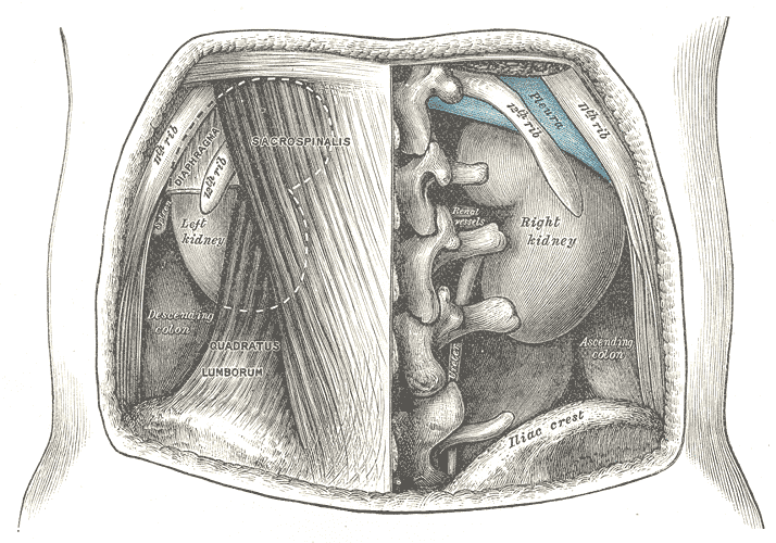
There are four organs and only the first does anything complicated. The two kidneys are retroperitoneal, lying against the posterior abdominal wall at roughly the level of vertebrae T12 to L3, with the right sitting slightly lower because the liver is above it. Each is about 11 centimetres long, wrapped in a fibrous capsule and a protective cushion of perirenal fat, and each has an outer cortex and an inner medulla arranged into eight to eighteen pyramids whose tips, the papillae, drain into minor calyces, then major calyces, then the renal pelvis. Blood arrives through the renal arteries, which together carry around 20 to 25 per cent of cardiac output — far more than the organ needs for its own metabolism, because it is there to be filtered rather than fed.
The ureters are muscular tubes about 25 centimetres long carrying urine from each renal pelvis to the bladder by peristalsis, not gravity, which is why lying down does not stop urine flow. They narrow at three points — the pelviureteric junction, where they cross the pelvic brim, and where they enter the bladder — and those three are exactly where stones lodge. They enter the bladder obliquely through its wall, an arrangement that acts as a valve: as the bladder fills, its own pressure compresses the intramural ureter and prevents reflux back toward the kidney.
The bladder is a hollow muscular reservoir in the pelvis, lined by transitional epithelium and walled by the detrusor, a mesh of smooth muscle running in several directions. Its interior is mostly folded into rugae that flatten as it fills, except at the trigone, a smooth triangle between the two ureteric orifices and the internal urethral opening, which stays flat at all volumes. Two sphincters guard the outlet: an internal sphincter of smooth muscle under autonomic control, and an external sphincter of skeletal muscle under voluntary control. The urethra then differs by sex — about 4 centimetres in women and around 20 in men, where it also carries semen — and that single difference explains most of the epidemiology of urinary infection.
How the system works
Urine formation is filtration followed by selective recovery. Blood pressure in the glomerular capillaries, much higher than in an ordinary capillary bed because the vessel on each side is an arteriole, pushes fluid across a three-layered barrier of fenestrated endothelium, thick basement membrane and podocyte filtration slits. Cells and plasma proteins stay behind, both because of size and because the membrane carries a fixed negative charge repelling albumin. The result is a glomerular filtration rate of roughly 125 millilitres per minute — 180 litres a day, from a blood volume of five. More than 99 per cent is then reabsorbed, so filtering everything and choosing afterwards is not wasteful but is what allows continuous regulation.
Each tubule segment does a different job. The proximal tubule reclaims about two-thirds of the water and sodium and essentially all the glucose, amino acids and bicarbonate. The loop of Henle builds a salt gradient in the medulla by countercurrent multiplication, so that the collecting duct passing back through it can pull water out of the urine when needed. The distal tubule and collecting duct fine-tune everything under hormonal control: aldosterone increases sodium reabsorption and potassium secretion, antidiuretic hormone inserts aquaporin channels that let water leave, and parathyroid hormone adjusts calcium. The tubule also secretes, actively adding hydrogen ions, potassium, creatinine and many drugs into the filtrate.
Beyond waste, four regulatory jobs matter clinically. The kidney controls extracellular fluid volume and therefore long-term blood pressure, through sodium handling and the renin-angiotensin-aldosterone system — which is why most antihypertensive drugs act on the kidney or its hormones. It controls acid-base balance by reabsorbing bicarbonate and excreting hydrogen ions, the only route by which the body can excrete fixed acid at all. It releases erythropoietin in response to low oxygen, which is why chronic kidney disease causes anaemia. And it performs the final hydroxylation activating vitamin D, which is why kidney failure causes bone disease. Emptying is a separate reflex: stretch receptors in the bladder wall signal at around 300 to 400 millilitres, a spinal reflex contracts the detrusor and relaxes the internal sphincter, and higher centres either permit or postpone this by controlling the external sphincter — voluntary control that is learned in childhood and lost with spinal cord injury.
The cells that do the work
Each segment is lined by a cell built for its specific job, and the histology follows the physiology exactly. Proximal tubule cells are the workhorses: tall cuboidal, with a dense apical brush border of microvilli multiplying absorptive surface, enormous numbers of mitochondria in deep basolateral infoldings to power the sodium pumps, and endocytic machinery to retrieve escaped protein. That metabolic demand, combined with sitting at the end of a long low-oxygen blood supply, is exactly why they die first in shock and are the classic site of acute tubular necrosis. The thin descending limb is simple squamous with few mitochondria because it only lets water leave passively. The thick ascending limb is mitochondria-rich to run the NKCC2 transporter that loop diuretics block. The collecting duct holds two populations: principal cells carrying the aldosterone and antidiuretic hormone receptors, and intercalated cells that secrete hydrogen or bicarbonate and are the final arbiter of blood pH. Podocytes deserve their own note: terminally differentiated, unable to divide, and once lost not replaced — which is the central reason glomerular damage tends to be permanent while tubular damage often is not. The bladder's urothelium solves a different problem, its surface umbrella cells joined by exceptionally tight junctions and coated with uroplakin plaques that make it the most impermeable epithelium in the body.
Under the microscope
Renal cortex is one of the most recognisable slides in histology. Renal corpuscles appear as balls of capillaries in a clear Bowman's space, and scattered between them are two kinds of cut tubule that students are asked to separate constantly. Proximal convoluted tubules are more numerous because they are longer and more convoluted, and they have a ragged lumen filled by a fuzzy pink brush border, deeply eosinophilic cytoplasm from the mitochondria, and few nuclei in any one profile because the cells are large. Distal convoluted tubules have a clean wide lumen with no brush border, paler cytoplasm and more nuclei per cross-section. Where a distal tubule touches its own glomerulus its cells crowd into the macula densa. The medulla looks entirely different: parallel straight tubules and collecting ducts running in one direction, with thin limbs lined by flat cells hard to distinguish from the vasa recta beside them. The ureter and bladder share transitional epithelium, recognised by large rounded dome-shaped surface cells, some binucleate, that flatten when the organ is distended — and the ureter adds a distinctive star-shaped lumen in cross-section.
When the system fails
Kidney failure is divided by time course because the causes and reversibility differ completely. Acute kidney injury develops over hours to days and is classified by where the problem sits: pre-renal, from reduced perfusion in dehydration, haemorrhage or sepsis, which is reversible if caught early; renal, from damage to the tissue itself, most often acute tubular necrosis from prolonged hypotension or nephrotoxic drugs; and post-renal, from obstruction. Chronic kidney disease develops over years, most commonly from diabetes and hypertension, and brings anaemia from lost erythropoietin, bone disease from failed vitamin D activation, acidosis, hyperkalaemia and fluid overload.
Glomerular disease presents in two patterns worth telling apart. Nephrotic syndrome is a permeability problem: heavy proteinuria, low plasma albumin, generalised oedema and high cholesterol, following podocyte injury. Nephritic syndrome is an inflammatory problem: haematuria, hypertension, reduced urine output and mild oedema, as in post-streptococcal glomerulonephritis and IgA nephropathy. Urinary tract infection is far commoner than either and is much more frequent in women, because a 4-centimetre urethra opening close to the anus is a short route for bowel organisms; infection confined to the bladder causes frequency, urgency and burning, whereas fever, rigors and loin pain mean it has reached the kidney.
Obstruction and storage problems complete the picture. Kidney stones cause colicky loin-to-groin pain as the ureter contracts against them, and they lodge at the three natural narrowings; most are calcium oxalate, and small ones pass spontaneously. In men, benign prostatic hyperplasia compresses the urethra, causing hesitancy, poor stream and incomplete emptying, and if unrelieved can back pressure up to the kidneys. Incontinence divides usefully into stress incontinence, leakage on coughing or lifting from a weak pelvic floor, and urge incontinence from an overactive detrusor — and the two are treated in entirely different ways, which is why the distinction is worth making before prescribing anything.
Common questions about the urinary system
What are the organs of the urinary system?
Two kidneys, two ureters, the bladder and the urethra. The kidneys filter blood and make urine, doing all the regulatory work; the ureters carry urine to the bladder by peristalsis; the bladder stores it; and the urethra carries it out. Only the kidney does anything metabolically complex — the other three are transport and storage.
What does the kidney do besides making urine?
Four things that matter as much as excretion. It controls extracellular fluid volume and therefore long-term blood pressure, largely through sodium handling and the renin-angiotensin-aldosterone system. It regulates blood pH, being the only route for excreting fixed acid. It releases erythropoietin to drive red cell production, which is why kidney failure causes anaemia. And it performs the final activation of vitamin D, which is why kidney failure causes bone disease.
How is urine made?
In three steps. Filtration: blood pressure in the glomerulus pushes water and small solutes into Bowman's capsule, about 180 litres a day. Reabsorption: the tubules reclaim more than 99 per cent of that, including all the glucose and amino acids and most of the water and salt. Secretion: the tubules actively add hydrogen ions, potassium, creatinine and many drugs into the filtrate. What is left, one to two litres, is urine.
Why are urinary tract infections more common in women?
Mainly anatomy. The female urethra is roughly 4 centimetres long compared with about 20 in males, and its opening lies close to the anus, so bowel organisms have a much shorter route to the bladder. Sexual activity, some contraceptives and the fall in oestrogen after the menopause add further risk. Infection confined to the bladder causes burning, frequency and urgency; fever, rigors and loin pain suggest it has reached the kidney and needs urgent treatment.
Selected references
- Hall JE, Hall ME. Guyton and Hall Textbook of Medical Physiology. 14th ed. Elsevier; 2021.
- Standring S, ed. Gray's Anatomy: The Anatomical Basis of Clinical Practice. 42nd ed. Elsevier; 2021.
- Ross MH, Pawlina W. Histology: A Text and Atlas. 8th ed. Wolters Kluwer; 2020.
- Levey AS, Inker LA, Coresh J. GFR estimation: from physiology to public health. American Journal of Kidney Diseases. 2014;63(5):820-834.
- Fowler CJ, Griffiths D, de Groat WC. The neural control of micturition. Nature Reviews Neuroscience. 2008;9(6):453-466.
- Kumar V, Abbas AK, Aster JC. Robbins and Cotran Pathologic Basis of Disease. 10th ed. Elsevier; 2021.
Medical disclaimer. This article is written for education and general understanding. It is not medical advice and cannot replace assessment by a qualified healthcare professional. If you have symptoms or health concerns, speak with a clinician. Read the full disclaimer.