Every joint in the body is solving the same problem twice: hold two bones together, and let them move. Those goals fight each other, and the skeleton's answer is to pick a different point on the trade-off in every location. The sutures of the skull give up all movement for absolute stability; the shoulder gives up stability for the widest range of motion of any joint in the body, and dislocates more often than any other as a direct result. Learning joints is really learning to read that compromise off the anatomy.
Where it is, and what it looks like
The structural classification asks what physically lies between the two bones. In a fibrous joint, dense connective tissue binds the bones directly with no cavity between them: the sutures of the skull, the syndesmoses that tie the radius to the ulna and the tibia to the fibula, and the gomphoses that hold each tooth in its socket. In a cartilaginous joint, cartilage bridges the gap. Primary cartilaginous joints, or synchondroses, use hyaline cartilage and are usually temporary, as at the epiphyseal growth plates; secondary cartilaginous joints, or symphyses, use fibrocartilage and are permanent, as at the intervertebral discs and pubic symphysis. In a synovial joint the bones do not touch at all, being separated by a fluid-filled cavity.
The functional classification asks instead how much movement is allowed, and it maps onto the structural one only loosely. Synarthroses are immovable, amphiarthroses are slightly movable, and diarthroses move freely. Most fibrous joints are synarthroses and most cartilaginous joints are amphiarthroses, but there are exceptions in both directions, which is exactly why both classifications are kept. Every synovial joint is a diarthrosis.
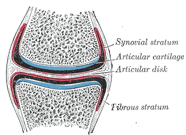
Synovial joints share a standard set of parts. Articular cartilage, usually hyaline, caps each bone end. A fibrous capsule encloses the joint and is lined on its inner surface by synovial membrane, which secretes synovial fluid into the cavity. Ligaments reinforce the capsule, either as thickenings within it or as separate structures inside or outside it. Many joints add accessory structures: menisci and articular discs that improve the fit between mismatched surfaces, labra that deepen a socket, fat pads that fill dead space, and bursae that reduce friction where tendons cross bone. Synovial joints are then subdivided into six shapes — plane, hinge, pivot, condyloid, saddle and ball-and-socket — in ascending order of freedom.
What it does
A synovial joint moves in one, two or three planes, and the shape of its surfaces decides which. Plane joints, as between the carpal bones, slide. Hinge joints such as the elbow, knee and interphalangeal joints allow flexion and extension in one plane only. Pivot joints such as the atlantoaxial joint and the proximal radioulnar joint allow rotation about a single axis. Condyloid joints such as the wrist and the metacarpophalangeal joints move in two planes, giving flexion, extension, abduction, adduction and circumduction but no true rotation. Saddle joints, of which the thumb's carpometacarpal joint is the important example, do the same with much greater freedom and are what makes the human thumb oppose. Ball-and-socket joints at the hip and shoulder move in all three planes.
Synovial fluid does three jobs and is made from two ingredients. It is a dialysate of blood plasma with hyaluronan and lubricin added by the synovial membrane, and it lubricates the surfaces, absorbs shock, and delivers nutrients to the avascular articular cartilage while removing waste. It is also thixotropic: it becomes less viscous as shear rate rises, so a joint that is moving quickly is better lubricated than one at rest. That behaviour explains the everyday experience of joints feeling stiff after inactivity and easing with movement, and it is one reason exercise protects cartilage rather than wearing it out.
Stability comes from four sources, and joints differ in which they rely on. Bony congruence — how well the surfaces fit — is the strongest, and it dominates at the hip, where a deep acetabulum makes dislocation rare and requires major force. Ligaments provide passive restraint at the end of range. Muscles and their tendons provide active, adjustable restraint, and at the shoulder they are the main mechanism, since the glenoid fossa is shallow and the rotator cuff does most of the holding. Negative intra-articular pressure adds a small suction effect. The general rule is that stability and mobility trade against each other, and where mobility wins, the joint depends on muscle — which is why rehabilitation after a shoulder injury is largely muscular.
The cells that build it
The tissues meeting at a synovial joint are an unusual combination, because two of them have no blood supply and one has no nerves. Articular cartilage is hyaline cartilage without a perichondrium, avascular, aneural and alymphatic, fed entirely by diffusion from synovial fluid driven by cyclical loading. It contains no nerve endings at all, which is why cartilage damage is painless and why joint pain arises instead from the subchondral bone, the fibrous capsule, the ligaments and above all the synovium, all of which are richly innervated. The synovial membrane is not an epithelium despite lining a cavity: it has no basement membrane and consists of one to three layers of two cell types, type A synoviocytes, which are macrophage-derived and phagocytose debris, and type B synoviocytes, which are fibroblast-like and secrete hyaluronan and lubricin. Beneath them lies a vascular subintima that supplies the fluid. Joints are also densely supplied with proprioceptors — Ruffini endings, Pacinian corpuscles and Golgi tendon organs in the capsule and ligaments — which report joint position and tension continuously to the spinal cord and brain. Hilton's law captures the arrangement neatly: the nerve supplying a muscle that moves a joint also supplies the joint itself and the skin over it, which is why joint pathology so often refers pain to a distant patch of skin.
How to recognise it on a slide
A section through a synovial joint shows a sequence of tissues in a fixed order, and reading it outward from the cavity is the easiest approach. The joint space itself appears empty because the fluid is washed out during processing. Facing it on each side is articular cartilage: a smooth pale matrix with chondrocytes in lacunae, flattened and parallel to the surface in the superficial zone, rounded and in isogenous groups in the middle zone, and arranged in vertical columns in the deep zone, separated from the calcified layer beneath by a wavy basophilic line called the tidemark. Below that, calcified cartilage merges into subchondral bone. Lining the rest of the cavity is the synovial membrane, distinguishable from epithelium precisely by what it lacks: no basement membrane, no tight junctions, and an irregular one-to-three-cell layer whose surface cells are not joined into a continuous sheet, often thrown into villi and resting on vascular loose connective tissue or fat. Outside that lies the fibrous capsule, dense irregular connective tissue continuous with the periosteum of both bones, and blending into it the ligaments, which are dense regular connective tissue with their collagen running in parallel waves.
When it goes wrong
Osteoarthritis is the mechanical failure of a joint and the commonest joint disease worldwide. Articular cartilage thins and fissures, the subchondral bone thickens and forms cysts, osteophytes grow at the joint margins, and the synovium becomes mildly inflamed. It typically affects the knees, hips, hands and spine, is worse with use and better with rest, and produces stiffness that lasts under thirty minutes in the morning. Rheumatoid arthritis is a different disease with a different target: an autoimmune inflammation of the synovium itself, which proliferates into an invasive pannus that erodes cartilage and bone from the joint margins inward. It is symmetrical, favours the small joints of the hands and feet, and causes morning stiffness lasting more than an hour.
Acute joint problems are dominated by three. A dislocation is complete loss of contact between the articular surfaces and reflects the mobility-stability trade-off directly — the shoulder accounts for around half of all dislocations because it is the most mobile joint in the body. Ligament sprains are graded by whether the fibres are stretched, partially torn or completely ruptured, and the anterior cruciate ligament of the knee heals poorly because it sits inside the joint bathed in synovial fluid, away from the blood supply a healing ligament needs. Meniscal tears heal only in the outer third, the only region that is vascularised.
Two further categories are worth recognising quickly. Septic arthritis is infection within the joint space, and it destroys cartilage within days through bacterial enzymes and the host inflammatory response; a hot, swollen, exquisitely painful joint that the patient will not move, with fever, is an emergency requiring aspiration and antibiotics rather than observation. Crystal arthropathies produce a similar picture: gout, from monosodium urate crystals, classically strikes the first metatarsophalangeal joint, while pseudogout, from calcium pyrophosphate, favours the knee and wrist. Polarised light microscopy of aspirated fluid separates all three, which is why joint aspiration remains the single most useful investigation in an acutely inflamed joint.
Common questions about the joints
What are the three main types of joints?
Structurally, joints are fibrous, cartilaginous or synovial. Fibrous joints are held by dense connective tissue with no cavity, as in skull sutures and the tooth socket. Cartilaginous joints are bridged by cartilage, either hyaline at growth plates or fibrocartilage at intervertebral discs and the pubic symphysis. Synovial joints have a fluid-filled cavity between the bones and are the freely movable joints of the limbs.
What are the six types of synovial joint?
Plane joints slide, as between the carpal bones. Hinge joints move in one plane, as at the elbow and knee. Pivot joints rotate about one axis, as at the atlantoaxial and proximal radioulnar joints. Condyloid joints move in two planes, as at the wrist and knuckles. Saddle joints move in two planes with greater freedom, as at the base of the thumb. Ball-and-socket joints move in all three, as at the hip and shoulder.
What does synovial fluid do?
Three things. It lubricates the joint, using hyaluronan and lubricin secreted by the synovial membrane, to a degree that gives less friction than ice on ice. It absorbs and distributes load across the cartilage surface. And it feeds the articular cartilage, which has no blood supply of its own, delivering nutrients and removing waste as the fluid is pumped in and out of the matrix by movement.
Why is the shoulder dislocated more often than the hip?
Because they sit at opposite ends of the same trade-off. The hip is a deep ball-and-socket joint whose bony socket encloses most of the femoral head, so it is stable and needs major force to dislocate. The shoulder's socket, the glenoid fossa, is shallow and covers only a small part of the humeral head, which buys an enormous range of motion at the cost of bony stability. It relies on the rotator cuff muscles instead, which is why shoulder rehabilitation is mostly muscular.
Selected references
- Standring S, ed. Gray's Anatomy: The Anatomical Basis of Clinical Practice. 42nd ed. Elsevier; 2021.
- Drake RL, Vogl AW, Mitchell AWM. Gray's Anatomy for Students. 4th ed. Elsevier; 2020.
- Ross MH, Pawlina W. Histology: A Text and Atlas. 8th ed. Wolters Kluwer; 2020.
- Sophia Fox AJ, Bedi A, Rodeo SA. The basic science of articular cartilage: structure, composition, and function. Sports Health. 2009;1(6):461-468.
- Smith MD. The normal synovium. The Open Rheumatology Journal. 2011;5:100-106.
- Kumar V, Abbas AK, Aster JC. Robbins and Cotran Pathologic Basis of Disease. 10th ed. Elsevier; 2021.
Medical disclaimer. This article is written for education and general understanding. It is not medical advice and cannot replace assessment by a qualified healthcare professional. If you have symptoms or health concerns, speak with a clinician. Read the full disclaimer.