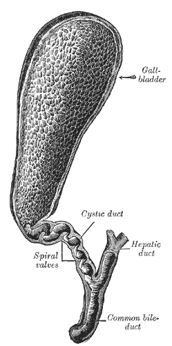
The gallbladder is the smallest organ most people can name, and the one most often removed. It holds barely 50 millilitres, yet it sits downstream of a liver that produces up to a litre of bile a day, and it manages that mismatch by concentrating what it stores up to tenfold. Reading it properly means following the bile: where it is made, where it is parked, what the wall does to it while it waits, and what releases it into the duodenum at exactly the moment fat arrives.
Anatomy: the structure
The gallbladder is a pear-shaped sac roughly 7 to 10 cm long, lying in a shallow fossa on the visceral surface of the liver between the right and quadrate lobes, along the line that separates them. It is described in three parts. The fundus is the rounded blind end that projects just past the inferior border of the liver and meets the anterior abdominal wall where the ninth costal cartilage crosses the lateral edge of the rectus sheath, which is the surface marking used when eliciting Murphy sign. The body runs backwards and upwards in contact with the liver and the first part of the duodenum, and the neck narrows into the cystic duct after an eccentric outpouching known as the infundibulum or Hartmann pouch, which is where stones commonly lodge.
The biliary tree drains into it and out of it. The right and left hepatic ducts join at the porta hepatis to form the common hepatic duct, which is joined by the cystic duct to form the common bile duct. The cystic duct is only 3 to 4 cm long and its mucosa is thrown into a crescentic spiral fold, the spiral valve of Heister, which holds the lumen open rather than acting as a true valve. The common bile duct descends in the free edge of the hepatoduodenal ligament, to the right of the hepatic artery and anterior to the portal vein, passes behind the first part of the duodenum, grooves or tunnels through the head of the pancreas, and usually joins the main pancreatic duct to form the hepatopancreatic ampulla, which opens at the major duodenal papilla. Smooth muscle around that ampulla forms the sphincter of Oddi.
The arterial supply is the cystic artery, normally a branch of the right hepatic artery, and it is found within the cystohepatic triangle of Calot, bounded by the cystic duct, the common hepatic duct, and the inferior surface of the liver. Venous drainage is unusual: small veins pass directly from the gallbladder bed into the liver substance rather than collecting into one cystic vein, which is why bleeding from the liver bed is a separate surgical problem from bleeding from the artery. Lymph drains to the cystic node at the neck, then to hilar and coeliac nodes. Innervation comes from the coeliac plexus, the vagus, and the right phrenic nerve, and it is the phrenic contribution, carrying fibres from C3 to C5, that refers gallbladder pain to the tip of the right shoulder.
Physiology: how it works
The liver secretes 600 to 1,000 millilitres of bile each day, and its composition explains everything the gallbladder does. Bile is mostly water, with bile salts, phospholipid, cholesterol, conjugated bilirubin, proteins, and electrolytes. Bile salts are amphipathic, so above a critical concentration they assemble into micelles that carry cholesterol and the products of fat digestion in aqueous solution. Between meals the sphincter of Oddi is closed, pressure in the duct system rises above the pressure inside the gallbladder, and bile is diverted along the cystic duct into storage instead of into the duodenum.
Storage is not passive. The epithelium actively absorbs sodium through sodium-hydrogen exchange, with chloride following through chloride-bicarbonate exchange, and water follows the resulting osmotic gradient through aquaporin channels. The gallbladder therefore reduces the volume of what it holds five to tenfold while leaving the bile salts, cholesterol, and bilirubin behind, which is why gallbladder bile is dark, viscous, and far more concentrated than hepatic bile. The same process explains why the gallbladder is where cholesterol stones form: concentrating the solutes pushes cholesterol toward the limit of what the bile salt and lecithin micelles can hold in solution.
Emptying is driven by cholecystokinin. Fat and protein digestion products in the duodenum stimulate the I cells of the duodenal and jejunal mucosa to release cholecystokinin, which contracts gallbladder smooth muscle through CCK-1 receptors and relaxes the sphincter of Oddi, while vagal cholinergic input reinforces the contraction; roughly half to two-thirds of the stored volume is delivered over 30 to 40 minutes. What is released is largely recovered. About 95 percent of bile salts are reabsorbed in the terminal ileum by the apical sodium-dependent bile acid transporter, returned to the liver in portal blood, and secreted again, so a pool of only 3 to 4 grams recycles six to ten times a day. Bile acids arriving back at the hepatocyte activate the nuclear receptor FXR, which restrains further synthesis and closes the loop.
Biology: the living cells
The gallbladder epithelium is a single layer of tall columnar cells built for transport rather than for secretion of digestive enzymes. Each cell carries short apical microvilli, is sealed to its neighbours by tight junctions, and places the sodium-potassium ATPase along its basolateral membrane, so sodium taken up at the apical surface is pumped out into the lateral intercellular space and water follows. Aquaporins in both membranes make that water movement fast enough to concentrate bile between meals. Mucus-secreting cells cluster in the neck and produce the glycoprotein layer that shields the epithelium from the detergent action of concentrated bile salts, and the same mucin becomes a nucleating scaffold when stones form. The smooth muscle of the wall is an irregular meshwork rather than discrete layers, carries CCK-1 and muscarinic receptors, and is coupled by gap junctions so that contraction is coordinated across the whole organ. The cholangiocytes lining the ducts upstream are a different cell again: they express CFTR and the chloride-bicarbonate exchanger AE2 and add a bicarbonate-rich secretion under the influence of secretin, thinning and alkalinising bile as it travels. Across all of these cells bile acids act not only as detergents but as signalling molecules, through FXR in the nucleus and TGR5 at the membrane.
Histology: under the microscope
The gallbladder wall is the classic exception in the gastrointestinal tract, and identifying it under the microscope is largely a matter of noticing what is missing. The mucosa is a simple tall columnar epithelium thrown into deep, branching, interconnecting folds that resemble villi in section but are not, resting on a lamina propria of loose vascular connective tissue. There is no muscularis mucosae and no submucosa, so the lamina propria sits directly against the muscle. That muscle is a fibromuscular coat of smooth muscle bundles running in several directions, mixed with collagen and elastic fibres rather than organised into inner circular and outer longitudinal layers. Outside it lies a perimuscular connective tissue layer, covered by serosa on the free surface and blending into the liver capsule as adventitia where the organ is attached. Two named features complete the picture: Rokitansky-Aschoff sinuses are outpouchings of mucosa that herniate into and through the muscle layer, common in chronic inflammation, and the ducts of Luschka are small accessory bile ducts in the liver bed that can leak after cholecystectomy. There are no goblet cells in a normal gallbladder, and their appearance is a metaplastic change.
Clinical correlations
Gallstones form when the solubility limits described above are exceeded. Cholesterol stones, the majority in Western populations, need supersaturated bile, a nucleating factor such as mucin, and gallbladder stasis; the familiar risk factors of female sex, increasing age, obesity, and pregnancy all act through those three. Pigment stones instead reflect excess unconjugated bilirubin, from chronic haemolysis or from bacterial deconjugation in infected bile. A stone that intermittently obstructs the neck or cystic duct produces biliary colic, a constant right upper quadrant pain that builds after a fatty meal and settles over hours. Sustained obstruction produces acute cholecystitis, in which the wall becomes inflamed and oedematous, and inspiration during palpation of the right upper quadrant arrests as the tender fundus meets the examining hand.
Obstruction lower in the tree causes different problems. A stone in the common bile duct blocks conjugated bilirubin from reaching the gut, producing obstructive jaundice with pale stools, dark urine, itching, and a rise in conjugated bilirubin and alkaline phosphatase out of proportion to the transaminases. Stagnant obstructed bile becomes infected easily, and ascending cholangitis presents with the Charcot triad of fever, jaundice, and right upper quadrant pain. A stone impacted at the ampulla can obstruct the pancreatic duct as well, which is the mechanism of gallstone pancreatitis and the reason the shared ampullary anatomy matters clinically. Courvoisier reasoned that a palpable, painless, distended gallbladder alongside jaundice is unlikely to be caused by stones, because a chronically stone-diseased gallbladder is fibrotic and cannot distend.
Cholecystectomy is one of the most common abdominal operations, and its risks are anatomical. The critical view of safety in the triangle of Calot exists because the cystic and common bile ducts can be confused and because the cystic artery is variable; injury to the common bile duct is the serious complication. Mirizzi syndrome, in which a stone in the neck compresses the adjacent common hepatic duct, reproduces obstruction without the stone ever leaving the gallbladder. After removal, bile drains continuously into the duodenum at a low rate instead of arriving as a bolus, which most people tolerate without any change in diet, though a minority develop bile acid diarrhoea. Chronic irritation carries its own risk: long-standing stones and a heavily calcified porcelain gallbladder are associated with gallbladder carcinoma, an uncommon but aggressive tumour that spreads early into the adjacent liver bed.
Common questions about the gallbladder
What does the gallbladder actually do?
It stores and concentrates bile between meals, then delivers it in a bolus when food reaches the duodenum. It does not make bile; the liver does. The gallbladder absorbs sodium, chloride, and water from the bile it holds, which is why gallbladder bile is far more concentrated than the bile arriving from the liver.
Can you live normally without a gallbladder?
Yes. After cholecystectomy bile flows continuously from the liver into the duodenum at a lower rate instead of being released as a bolus, and the bile ducts dilate slightly to compensate. Most people notice no difference and need no special diet, although a minority develop looser stools because unabsorbed bile acids reach the colon.
How do you recognise the gallbladder under a microscope?
Look for a simple tall columnar epithelium with deep branching mucosal folds sitting on a lamina propria that lies directly against a disorganised fibromuscular coat, with no muscularis mucosae and no submucosa between them. Rokitansky-Aschoff sinuses, where mucosa herniates into the muscle, confirm it, and a normal gallbladder has no goblet cells.
Why do gallstones form?
Cholesterol stones need three things at once: bile supersaturated with cholesterol relative to the bile salts and lecithin available to hold it in micelles, a nucleating factor such as mucin, and stasis that gives crystals time to grow. Pigment stones form instead when unconjugated bilirubin is in excess, from haemolysis or from bacterial infection of bile.
Selected references
- Standring S, ed. Gray's Anatomy: The Anatomical Basis of Clinical Practice. 42nd ed. Elsevier; 2021.
- Ross MH, Pawlina W. Histology: A Text and Atlas. 8th ed. Wolters Kluwer; 2020.
- Hall JE, Hall ME. Guyton and Hall Textbook of Medical Physiology. 14th ed. Elsevier; 2021.
- Boyer JL. Bile formation and secretion. Comprehensive Physiology. 2013;3(3):1035-1078.
- Barrett KE, Barman SM, Brooks HL, Yuan JXJ. Ganong's Review of Medical Physiology. 26th ed. McGraw Hill; 2019.
- Kumar V, Abbas AK, Aster JC. Robbins and Cotran Pathologic Basis of Disease. 10th ed. Elsevier; 2021.
Medical disclaimer. This article is written for education and general understanding. It is not medical advice and cannot replace assessment by a qualified healthcare professional. If you have symptoms or health concerns, speak with a clinician. Read the full disclaimer.