Each adrenal gland weighs about four grams and sits like a cap on the upper pole of a kidney, and almost nothing about it is what it first appears. It is not one gland but two fused together, with different embryonic origins, different signals, and different products: an outer cortex making steroid hormones from cholesterol, and an inner medulla that is really a sympathetic ganglion which lost its axons and started secreting into blood instead. Understanding the adrenal means keeping those two halves separate, then seeing how the blood flowing from one into the other ties them back together.
Anatomy: the structure
The adrenal, or suprarenal, glands are retroperitoneal structures lying on the superomedial aspect of each kidney at about the level of the twelfth thoracic vertebra. They sit within the renal fascia but are separated from the kidney itself by a septum of perirenal fat, which is why a kidney that drops with age or after nephrectomy leaves its adrenal behind. The right gland is pyramidal and lies higher and more medially, wedged behind the inferior vena cava and against the bare area of the liver. The left is flatter and semilunar, related to the stomach, the pancreas, and the splenic vessels. Each measures roughly 5 cm by 3 cm by 1 cm and weighs 4 to 6 grams, and a cut surface shows a firm yellow cortex, coloured by stored lipid, around a thin dark red-brown medulla.
The blood supply is disproportionate to the size of the organ and is arranged as an inflow from three sources and an outflow through one vein. Superior suprarenal branches come from the inferior phrenic artery, middle suprarenal branches arise directly from the abdominal aorta, and inferior suprarenal branches come from the renal artery; together they form a subcapsular plexus feeding as many as sixty small vessels into the gland. Venous drainage is asymmetrical and surgically important: the right suprarenal vein is short and drains straight into the inferior vena cava, while the left is longer and drains into the left renal vein. Lymphatics follow to the lumbar para-aortic nodes.
The two halves have separate origins. The cortex arises from coelomic mesoderm near the urogenital ridge, which is why adrenal and gonadal steroidogenesis share so much machinery, and the fetus builds a large fetal cortex that involutes over the first year of life. The medulla is colonised by neural crest cells that migrate in and differentiate into chromaffin cells, so it is developmentally a modified sympathetic ganglion. That origin dictates its innervation: preganglionic sympathetic fibres from the greater splanchnic nerve, carrying segments T5 to T9, pass through the coeliac plexus and synapse directly on chromaffin cells, with no postganglionic neuron in between. Arterial blood also drains inward through cortical sinusoids before reaching the medulla, so medullary cells are bathed in blood carrying very high concentrations of cortical steroid.
Physiology: how it works
The cortex makes three classes of steroid, and they are laid out in order from outside in: mineralocorticoids from the zona glomerulosa, glucocorticoids from the zona fasciculata, and androgen precursors from the zona reticularis. All of them start from cholesterol, and the rate-limiting step in every zone is the transfer of cholesterol into the mitochondrion by the StAR protein, followed by side-chain cleavage by CYP11A1 to pregnenolone. What differs between the zones is which downstream enzymes they express. Only the glomerulosa carries aldosterone synthase, CYP11B2, so only it can complete aldosterone; the glomerulosa lacks CYP17A1, so it cannot make cortisol or androgens; and the reticularis expresses high CYP17A1 lyase activity with sulfotransferase, so it exports DHEA and DHEA sulfate.
The two main products are controlled by completely separate systems, which is the single most useful fact about adrenal physiology. Cortisol is governed by the hypothalamic-pituitary-adrenal axis: corticotropin-releasing hormone drives pituitary ACTH, cleaved from proopiomelanocortin, and ACTH drives the fasciculata and reticularis; cortisol then feeds back negatively on both. Output follows a diurnal rhythm peaking in the early morning and rises sharply with physical and psychological stress. Cortisol raises blood glucose by promoting gluconeogenesis, proteolysis, and lipolysis, is permissive for the vascular action of catecholamines, suppresses inflammation and immunity at high concentrations, and inhibits bone formation. It binds the mineralocorticoid receptor as tightly as aldosterone does, and the kidney only escapes being flooded with a mineralocorticoid signal because 11-beta-hydroxysteroid dehydrogenase type 2 converts cortisol to inactive cortisone in the tubular cells.
Aldosterone answers to the renin-angiotensin system and to plasma potassium, not to ACTH; a fall in renal perfusion pressure or in sodium delivery releases renin, angiotensin II is generated, and both angiotensin II and a rising potassium stimulate the glomerulosa directly. Aldosterone then acts on principal cells of the collecting duct to insert epithelial sodium channels and sodium-potassium ATPase, so sodium and water are retained while potassium and hydrogen ions are excreted. The medulla runs on a different timescale entirely. Tyrosine is converted to DOPA by tyrosine hydroxylase, the rate-limiting enzyme, then to dopamine, then to noradrenaline, and finally to adrenaline by PNMT, an enzyme induced by the very high cortisol concentrations arriving from the cortex, which is why adrenaline is made almost nowhere else. Roughly 80 percent of what is stored and released is adrenaline. Splanchnic stimulation triggers exocytosis of the granules, and because the hormones then travel in blood rather than across a synapse their effect lasts about ten times longer than direct sympathetic nerve activity. Catecholamines are broken down by monoamine oxidase and catechol-O-methyltransferase to metanephrines and finally vanillylmandelic acid.
Biology: the living cells
Every steroid-secreting cell in the cortex carries the same structural signature: abundant smooth endoplasmic reticulum, mitochondria with tubular or vesicular rather than shelf-like cristae, and lipid droplets holding cholesterol esters. This reflects a synthetic pathway that shuttles intermediates back and forth between mitochondrion and smooth endoplasmic reticulum rather than running in one compartment. Steroids are not stored in granules, so secretion depends on synthesis, and output can only rise as fast as the cell can make new hormone. The zones differ in detail. Glomerulosa cells are small, relatively lipid-poor, and packed into rounded clusters. Fasciculata cells are large and so heavily loaded with lipid that routine processing dissolves it out and leaves them vacuolated, which is why they are called spongiocytes. Reticularis cells are smaller and darker, with less lipid and conspicuous lipofuscin from a long working life. Chromaffin cells of the medulla are modified postganglionic neurons without axons or dendrites; they store catecholamines in dense-core granules together with chromogranin A, ATP, and enkephalins, and they take their name from the brown colour that develops when chromium salts oxidise those catecholamines. They are supported by glial-like sustentacular cells that express S100, and scattered true sympathetic ganglion cells remain among them. The cortex renews itself from progenitor cells in the capsule and subcapsular region, whose descendants migrate inwards and change their phenotype as they pass from one zone to the next, which is why the zones are functional states rather than fixed populations.
Histology: under the microscope
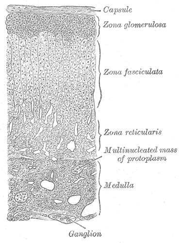
A low-power section shows four bands in order and their relative thickness is part of the identification. Beneath a dense collagenous capsule, the zona glomerulosa makes up around 15 percent of the cortex and is arranged in rounded, arch-shaped clusters of small cells. The zona fasciculata is much the largest at 65 to 80 percent, and its pale, vacuolated spongiocytes are stacked in straight cords one or two cells thick running radially toward the medulla, with long straight fenestrated sinusoids between them, giving a distinctive striped appearance at low power. The zona reticularis, about 7 to 10 percent, loses that order: the cords branch and anastomose into a network, and the cells are smaller, more eosinophilic, and often contain brown lipofuscin granules. The medulla is unmistakable once the cortex has been read, because the cells are large, pale, polyhedral, and grouped in clumps and short cords around wide venules rather than in radial cords, and because chromaffin granules stain with chromium salts. Two further details are diagnostic: the central medullary vein has an unusually thick, eccentric bundle of longitudinal smooth muscle in its wall, and the whole gland is drained by fenestrated sinusoids that make it one of the most vascular tissues in the body per unit mass.
Clinical correlations
Too much cortisol produces Cushing syndrome, with central obesity, a rounded plethoric face, purple striae, proximal muscle wasting, thin skin and easy bruising, glucose intolerance, hypertension, and osteoporosis. By far the commonest cause is prescribed glucocorticoid; of the endogenous causes, a pituitary corticotroph adenoma driving both glands is called Cushing disease, while a unilateral adrenal adenoma secretes independently and leaves ACTH suppressed and the other gland atrophic. Excess aldosterone from a glomerulosa adenoma or from bilateral hyperplasia produces primary hyperaldosteronism, in which hypertension is combined with a low or low-normal potassium and a suppressed plasma renin, and it is one of the few genuinely curable causes of high blood pressure.
Too little is more dangerous. Primary adrenal insufficiency, or Addison disease, destroys the whole cortex, most often by autoimmune adrenalitis in high-income countries and by tuberculosis elsewhere, so cortisol and aldosterone are lost together. The result is fatigue, weight loss, postural hypotension, hyponatraemia, hyperkalaemia, and hyperpigmentation of skin creases and buccal mucosa, because the same proopiomelanocortin precursor that yields the greatly increased ACTH also yields melanocyte-stimulating hormone. Secondary insufficiency, from pituitary disease or from abruptly stopping long-term steroids, spares aldosterone, since the renin-angiotensin system is intact, and causes no pigmentation, since ACTH is low. Either can decompensate into an adrenal crisis with shock during intercurrent illness, and Waterhouse-Friderichsen syndrome describes the acute bilateral haemorrhagic destruction that can accompany meningococcal sepsis.
Two more disorders follow directly from the biology. Congenital adrenal hyperplasia is a group of enzyme defects, of which 21-hydroxylase deficiency accounts for about 90 percent; cortisol and often aldosterone cannot be completed, negative feedback is lost, ACTH rises and hypertrophies both glands, and the accumulating precursors are shunted down the androgen pathway, producing virilisation with salt wasting in the severe form. Phaeochromocytoma is a chromaffin cell tumour of the medulla, classically described by the rule of tens and presenting with episodic headache, palpitations, sweating, and paroxysmal hypertension; it is diagnosed by measuring plasma or urinary metanephrines rather than the catecholamines themselves, and alpha blockade must always precede beta blockade so that unopposed alpha stimulation does not precipitate a hypertensive crisis. In children the equivalent neural crest tumour is neuroblastoma. Incidental adrenal masses found on imaging are common and are assessed for both hormone secretion and malignant features.
Common questions about the adrenal glands
What are the three zones of the adrenal cortex and what does each one make?
From outside in: the zona glomerulosa makes mineralocorticoids, mainly aldosterone, which controls sodium and potassium; the zona fasciculata makes glucocorticoids, mainly cortisol; and the zona reticularis makes androgen precursors such as DHEA. A common memory aid is salt, sugar, and sex, in that order from the capsule inwards.
What is the difference between the adrenal cortex and the adrenal medulla?
They are effectively two organs in one capsule. The cortex comes from mesoderm, makes steroid hormones from cholesterol, and is controlled by ACTH and by the renin-angiotensin system. The medulla comes from neural crest, makes catecholamines, and is controlled directly by preganglionic sympathetic fibres, so it behaves like a sympathetic ganglion that secretes into blood.
How can I tell the adrenal zones apart under a microscope?
Work inwards from the capsule and use both the pattern and the thickness. Glomerulosa is a thin outer band of small cells in rounded clusters. Fasciculata is by far the thickest zone, with pale vacuolated cells in straight radial cords separated by long sinusoids. Reticularis is a thin darker band where the cords branch into a network. The medulla is separate again: large pale polyhedral cells in clumps around venules.
What happens if the adrenal glands stop working?
Losing the whole cortex removes both cortisol and aldosterone, causing fatigue, weight loss, low blood pressure on standing, low sodium, high potassium, and darkening of skin creases from the raised ACTH. If the failure is instead due to pituitary disease or to stopping steroid treatment suddenly, aldosterone is preserved and there is no pigmentation. Both forms can decompensate into an adrenal crisis, which is a medical emergency.
Selected references
- Standring S, ed. Gray's Anatomy: The Anatomical Basis of Clinical Practice. 42nd ed. Elsevier; 2021.
- Ross MH, Pawlina W. Histology: A Text and Atlas. 8th ed. Wolters Kluwer; 2020.
- Hall JE, Hall ME. Guyton and Hall Textbook of Medical Physiology. 14th ed. Elsevier; 2021.
- Miller WL, Auchus RJ. The molecular biology, biochemistry, and physiology of human steroidogenesis and its disorders. Endocrine Reviews. 2011;32(1):81-151.
- Melmed S, Auchus RJ, Goldfine AB, Koenig RJ, Rosen CJ, eds. Williams Textbook of Endocrinology. 14th ed. Elsevier; 2020.
- Kumar V, Abbas AK, Aster JC. Robbins and Cotran Pathologic Basis of Disease. 10th ed. Elsevier; 2021.
Medical disclaimer. This article is written for education and general understanding. It is not medical advice and cannot replace assessment by a qualified healthcare professional. If you have symptoms or health concerns, speak with a clinician. Read the full disclaimer.