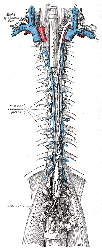
Capillaries leak. That is not a fault but a consequence of how exchange works: pressure pushes fluid out at the arterial end and osmotic pull returns most but not all of it, leaving around three litres a day stranded in the tissues. Without a way back, you would swell until circulation failed. The lymphatic system is that way back — a one-way network with no pump of its own that collects the surplus, carries it through a chain of filters where the immune system inspects everything passing through, and empties it into the veins at the base of the neck. Drainage and immunity in one set of vessels, which is why an infection makes the glands in your neck swell.
The parts, and how they fit together
The vessels begin as lymphatic capillaries, blind-ended tubes woven through almost every tissue. They are built for uptake rather than exchange: their endothelial cells overlap loosely to form flap-like valves that open inward when interstitial pressure rises, and they are anchored to surrounding tissue so that swelling pulls them open rather than collapsing them. That lets them admit not only fluid and protein but whole cells and bacteria, which blood capillaries cannot. They drain into collecting vessels with smooth muscle and abundant valves, then into larger trunks.
All of it converges on two ducts. The thoracic duct begins at the cisterna chyli in the abdomen and drains the entire lower body, the left side of the head, neck and chest, and the left arm — roughly three-quarters of the body — emptying into the junction of the left subclavian and internal jugular veins. The much smaller right lymphatic duct drains the right side of the head, neck and chest and the right arm into the equivalent point on the right. The asymmetry is embryological rather than functional, but it is clinically useful: a hard node just above the left clavicle can be the first sign of an abdominal cancer that has travelled the thoracic duct.
The organs divide into primary and secondary by what happens in them. Primary lymphoid organs are where lymphocytes are made and tested: the bone marrow, where all lymphocytes originate and B cells mature, and the thymus, prominent in childhood and shrinking after puberty, where T cells mature and most are destroyed for reacting to the body's own molecules. Secondary lymphoid organs are where lymphocytes meet antigen: hundreds of lymph nodes strung along the vessels; the spleen, which filters blood rather than lymph; the tonsils; and the mucosa-associated lymphoid tissue including the Peyer's patches of the ileum and the appendix.
How the system works
The first job is fluid balance, and the arithmetic is why it matters. Across all the capillaries in the body, filtration exceeds reabsorption by roughly 20 millilitres per minute, or about three litres a day — more than half the plasma volume. The lymphatics return that, and crucially they return the protein that leaked with it, which no other route can do; if protein accumulated in the interstitium it would draw yet more water out by osmosis and the swelling would accelerate. There is no pump, so flow depends entirely on external forces: skeletal muscle contraction squeezing vessels, arterial pulsation, respiratory pressure changes, and rhythmic contraction of the smooth muscle in the larger vessels, with valves ensuring everything moves one way.
The second job is fat absorption, and it is the reason a lymphatic vessel sits in the middle of every intestinal villus. Sugars and amino acids absorbed from the gut enter capillaries and go to the liver through the hepatic portal vein, but digested fat is reassembled inside the enterocyte into triglyceride, packaged as chylomicrons, and released into the lacteal instead. Chylomicrons are simply too large for a blood capillary. Lymph draining the gut after a meal is milky with them, which is what gives the cisterna chyli its name, and it reaches the bloodstream at the neck having bypassed the liver entirely.
The third job is immune surveillance, and the architecture is designed for it. Lymph from any tissue passes through at least one node before reaching the blood, entering through afferent vessels, percolating slowly through sinuses lined with macrophages, and leaving through a single efferent vessel. Anything the fluid picked up — bacteria, viruses, tumour cells, debris — is presented to the lymphocytes waiting there. Dendritic cells from the tissues also travel to the node deliberately, carrying antigen for presentation. Because a specific lymphocyte is rare and a specific antigen is rare, nodes exist to make that improbable meeting likely, and lymphocytes recirculate continuously through them. A node that becomes tender and swollen during infection is one where that process is running at full speed.
The cells that do the work
The cells are the same populations that run adaptive immunity, organised by geography. B lymphocytes recognise antigen directly in its native form and, once activated, become plasma cells secreting antibody; they occupy the follicles. T lymphocytes recognise only processed fragments displayed on MHC molecules, and split into CD8 cytotoxic cells that kill infected cells and CD4 helper cells that direct the response; they occupy the paracortex. Macrophages line the sinuses and phagocytose what passes. Dendritic cells are the couriers, sampling tissue, migrating to the node and presenting what they found. Follicular dendritic cells, a different lineage despite the name, hold antigen on their surface within germinal centres so B cells can compete for it. The supporting framework is reticular tissue, a mesh of type III collagen made by reticular cells, forming the scaffold that the wandering cells move along. Lymph itself is simply interstitial fluid that has entered a lymphatic — similar to plasma but with much less protein, and progressively enriched with lymphocytes as it passes through nodes, so efferent lymph carries far more cells than afferent lymph did.
Under the microscope
A lymph node is one of the most legible structures in histology because its compartments map exactly onto the two lymphocyte populations. Under a capsule, the outer cortex contains rounded follicles: a primary follicle is a small uniform ball of resting B cells, while a secondary follicle has a pale germinal centre of large dividing B cells, tingible-body macrophages containing the debris of apoptotic cells, and a darker mantle of small resting lymphocytes pushed to the edge. A germinal centre is direct visual evidence of an active immune response. The paracortex between and beneath the follicles is the T cell zone and expands in viral infections, while the follicles expand in bacterial ones. The medulla holds cords of plasma cells separated by sinuses draining toward the hilum. The spleen follows the same logic with different geography: white pulp forms sleeves around central arterioles, with the periarteriolar lymphoid sheath as the T zone and follicles budding off it as the B zone, all set in red pulp that filters blood and removes aged red cells. The thymus is unlike either, divided into lobules with a dark cortex packed with immature thymocytes, a paler medulla, and the distinctive concentric whorls of Hassall's corpuscles found nowhere else.
When the system fails
Lymphoedema is what happens when drainage fails, and it behaves unlike other swelling. Because the protein that leaked cannot be returned, it accumulates in the interstitium and holds water osmotically, so the swelling is persistent rather than fluctuating, does not pit readily once established, and progresses to fibrosis and skin thickening. The commonest cause worldwide is filariasis, a parasitic infection blocking the vessels; in high-income countries it most often follows surgery or radiotherapy that removed or scarred nodes, classically arm swelling after axillary node clearance for breast cancer. Treatment is compression, elevation and specialised massage rather than diuretics, which do not address the protein.
Enlarged nodes are one of the commonest reasons people seek medical advice, and the character matters more than the size. Reactive nodes from infection are tender, mobile, soft, and appear over days. Malignant nodes are typically painless, hard, fixed to surrounding tissue, and enlarge steadily over weeks. Generalised enlargement with fever, night sweats and weight loss raises the possibility of lymphoma, of which Hodgkin lymphoma is defined by the Reed-Sternberg cell on biopsy and the far more varied non-Hodgkin lymphomas are not. Because lymph flows in defined territories, the node group involved points back to the site of the problem, which is exactly why examining the draining nodes is part of examining any lesion.
The same pathways cancer uses for drainage it uses for spread, and that shapes surgery. Most carcinomas metastasise first to the nodes draining the primary tumour, which is why node status is central to staging and why the sentinel node — the first node in the chain — is biopsied to decide whether further clearance is needed. Losing the spleen has a specific consequence: it is a major site for responding to encapsulated bacteria, so patients without one need vaccination against pneumococcus, meningococcus and Haemophilus and often lifelong prophylactic antibiotics. Tonsils and adenoids, being the first lymphoid tissue air and food meet, enlarge in childhood and can obstruct breathing or the auditory tube, which is the usual reason for removing them.
Common questions about the lymphatic system
What does the lymphatic system do?
Three things. It returns roughly three litres a day of fluid and protein that leaks out of capillaries, without which tissues would swell continuously. It absorbs digested fat from the intestine as chylomicrons, bypassing the liver. And it houses the immune system's meeting places — lymph from every tissue passes through at least one node, where lymphocytes and macrophages inspect whatever it picked up.
What is the difference between lymph and blood?
Lymph is interstitial fluid that has entered a lymphatic vessel. It resembles plasma but contains much less protein, no red cells and no platelets, and it becomes progressively richer in lymphocytes as it passes through nodes. It moves in one direction only, from tissues toward the neck, and it has no pump — it depends on muscle contraction, breathing and valves, whereas blood circulates in a closed loop driven by the heart.
Why do lymph nodes swell when you are ill?
Because they are working. Antigen arriving in lymph, and dendritic cells carrying it from the infected tissue, trigger rapid proliferation of the specific lymphocytes that recognise it, so the node physically enlarges and its capsule stretches, which is what makes it tender. Reactive nodes are tender, soft and mobile, and settle. A node that is painless, hard, fixed and steadily growing over weeks is a different matter and needs assessment.
What causes lymphoedema and why does it not go away?
It follows blocked or removed lymphatics — most often filariasis worldwide, and surgery or radiotherapy to lymph nodes in high-income countries. It persists because the protein that leaked from capillaries cannot be returned by any other route, and that protein holds water osmotically in the tissue. Over time the swelling becomes fibrotic rather than fluid. This is why diuretics do not help and why treatment is compression, elevation and specialised drainage massage.
Selected references
- Standring S, ed. Gray's Anatomy: The Anatomical Basis of Clinical Practice. 42nd ed. Elsevier; 2021.
- Murphy K, Weaver C. Janeway's Immunobiology. 10th ed. W. W. Norton; 2022.
- Ross MH, Pawlina W. Histology: A Text and Atlas. 8th ed. Wolters Kluwer; 2020.
- Levick JR, Michel CC. Microvascular fluid exchange and the revised Starling principle. Cardiovascular Research. 2010;87(2):198-210.
- Alitalo K. The lymphatic vasculature in disease. Nature Medicine. 2011;17(11):1371-1380.
- Kumar V, Abbas AK, Aster JC. Robbins and Cotran Pathologic Basis of Disease. 10th ed. Elsevier; 2021.
Medical disclaimer. This article is written for education and general understanding. It is not medical advice and cannot replace assessment by a qualified healthcare professional. If you have symptoms or health concerns, speak with a clinician. Read the full disclaimer.