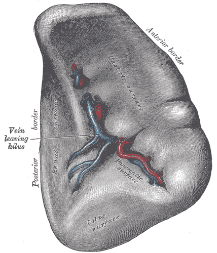
The spleen is the largest lymphoid organ in the body and the only one that filters blood rather than lymph. Roughly the size of a fist and weighing about 150 grams, it receives around 5 percent of cardiac output. Its job is quality control: every red blood cell must repeatedly squeeze through narrow slits in its walls, and cells too rigid to make the passage are removed. At the same time, its lymphoid compartments inspect blood-borne antigen.
Anatomy: the structure
The spleen lies in the left hypochondrium, along the axis of the ninth to eleventh ribs, and is normally impalpable because the rib cage covers it entirely; a spleen palpable below the costal margin has usually enlarged to two or three times its normal size. It is intraperitoneal except at the hilum and is supported by the gastrosplenic ligament, carrying the short gastric vessels, and the splenorenal ligament, carrying the splenic artery and vein and the tail of the pancreas.
Its visceral surface bears impressions from the structures it contacts: gastric, renal, colic, and pancreatic. The organ is enclosed by a capsule of dense connective tissue with some smooth muscle, and trabeculae extend inward from the capsule and hilum carrying vessels into the parenchyma. Because the capsule is thin and the parenchyma is soft and highly vascular, the spleen is the abdominal organ most often ruptured in blunt trauma, and its position under the lower left ribs makes fractures there a specific warning sign.
The splenic artery, the largest branch of the coeliac trunk, follows a characteristically tortuous course along the upper border of the pancreas before entering the hilum and dividing into segmental branches. Within the organ it becomes trabecular arteries, then central arterioles surrounded by lymphoid tissue, then penicillar arterioles opening into the red pulp. Venous blood collects into the splenic vein, which runs behind the pancreas and joins the superior mesenteric vein to form the portal vein, so portal hypertension is transmitted directly back to the spleen.
Physiology: how it works
Filtration depends on an unusual circulation. In the closed circulation, blood passes directly from arteriole to sinusoid; in the open circulation, which handles the majority of flow, blood is discharged into the splenic cords and must then re-enter the sinusoids by squeezing between endothelial cells through slits only 1 to 3 micrometres wide. A healthy erythrocyte, about 7 micrometres across, deforms and passes. An aged, antibody-coated, parasitised, or otherwise rigid cell cannot, and is retained and phagocytosed by cord macrophages.
Beyond removing whole cells, the spleen performs partial repairs through a process called pitting, in which macrophages remove inclusions such as Howell-Jolly bodies of nuclear remnant or Heinz bodies of denatured haemoglobin while leaving the cell intact and returning it to circulation. The appearance of Howell-Jolly bodies on a blood film is therefore a reliable indicator of absent or non-functioning splenic tissue. Iron recovered from degraded haemoglobin is returned to the transferrin pool for reuse.
Immunologically, the white pulp is the site where blood-borne antigen is presented. T lymphocytes occupy the periarteriolar lymphoid sheath around the central arteriole, B lymphocytes occupy adjacent follicles that develop germinal centres on activation, and the surrounding marginal zone contains specialised macrophages and B cells adapted to capture polysaccharide antigen. This last function explains the specific vulnerability created by splenectomy. The spleen also holds a reserve of about a third of the body's platelets and, in the fetus, is a site of haematopoiesis that can be reactivated in some diseases.
Biology: the living cells
Splenic function is built from cooperating cell populations arranged around blood flow. Cord macrophages in the red pulp express receptors for the Fc portion of antibody and for complement, letting them identify opsonised cells and particles, and they carry the enzymatic machinery for haem catabolism and iron recycling. Sinusoidal lining cells are elongated, arranged like the staves of a barrel with narrow gaps between them and a discontinuous basement membrane in hoops, which is precisely what forces erythrocytes to deform. Reticular cells and their reticulin fibres form the supporting meshwork of the cords. In the white pulp, follicular dendritic cells retain antigen for B cell selection, while marginal zone B cells mount rapid T-independent responses to encapsulated bacterial polysaccharide, generating IgM without requiring a full germinal centre reaction. This is why encapsulated organisms are the specific threat after splenectomy.
Histology: under the microscope
A low-power splenic section shows scattered pale nodules of white pulp set in a dark, diffusely red background of red pulp, a pattern no other organ reproduces. The diagnostic feature of white pulp is the central arteriole positioned eccentrically within or beside a lymphoid nodule, which distinguishes splenic follicles from those of a lymph node. Around the arteriole, the periarteriolar lymphoid sheath is T cell rich; the adjacent follicle is B cell rich; and the marginal zone forms the transitional band between white and red pulp where arterial blood is first discharged. The red pulp consists of splenic sinusoids, which are wide vessels with the characteristic gapped lining, separated by cords of Billroth packed with macrophages, plasma cells, and blood cells. Unlike a lymph node, the spleen has no cortex or medulla, no subcapsular sinus, and no afferent lymphatics, because its afferent supply is arterial blood rather than lymph.
Clinical correlations
Splenomegaly has causes that follow directly from splenic function. Portal hypertension in cirrhosis causes congestive enlargement because splenic venous drainage is portal. Haemolytic anaemias enlarge it through work hypertrophy as filtration demand rises. Infections such as infectious mononucleosis, malaria, and endocarditis enlarge it through immune activation, and infiltrative or haematological malignancies enlarge it directly. Hypersplenism describes the functional consequence: an overactive spleen sequestering cells and producing cytopenias.
Splenic rupture is a surgical emergency. Blunt trauma to the left upper quadrant, especially with lower rib fractures, can tear the capsule of an organ that receives 5 percent of cardiac output, producing rapid intraperitoneal haemorrhage. Referred pain to the left shoulder tip, the Kehr sign, occurs because blood irritates the diaphragm, which shares the C3 to C5 innervation of the shoulder.
After splenectomy or in functional asplenia such as sickle cell disease, patients lose marginal zone responses to polysaccharide antigen and become vulnerable to overwhelming post-splenectomy infection with encapsulated organisms, principally Streptococcus pneumoniae, Haemophilus influenzae type b, and Neisseria meningitidis. Vaccination against these before elective splenectomy and prompt treatment of febrile illness afterwards are standard practice. Blood film changes including Howell-Jolly bodies, target cells, and a rise in platelet count confirm the loss of splenic function.
Common questions about the spleen
What is the difference between red pulp and white pulp?
Red pulp is the filtering compartment, made of blood-filled sinusoids and the macrophage-rich cords of Billroth that remove aged or damaged red cells. White pulp is the immune compartment, made of lymphoid tissue arranged around central arterioles where blood-borne antigen is presented to T and B lymphocytes.
How does the spleen remove old red blood cells?
Most blood entering the spleen is discharged into the cords and must squeeze back into the sinusoids through slits far narrower than a red cell. Healthy flexible cells deform and pass, while aged or damaged cells that have lost membrane flexibility are held back and eaten by macrophages.
Can you live without a spleen?
Yes. The liver and bone marrow take over most filtering and recycling. However, the loss of marginal zone responses to bacterial capsular polysaccharide creates a lifelong risk of overwhelming infection with encapsulated organisms, so vaccination and rapid treatment of fever become important.
How do you distinguish spleen from lymph node histologically?
Look for a central arteriole inside the lymphoid nodule, which is unique to the spleen, and for the red pulp of sinusoids and cords surrounding it. A lymph node instead shows a distinct cortex and medulla, a subcapsular sinus, and afferent lymphatics, none of which the spleen has.
Selected references
- Standring S, ed. Gray's Anatomy: The Anatomical Basis of Clinical Practice. 42nd ed. Elsevier; 2021.
- Ross MH, Pawlina W. Histology: A Text and Atlas. 8th ed. Wolters Kluwer; 2020.
- Mebius RE, Kraal G. Structure and function of the spleen. Nature Reviews Immunology. 2005;5(8):606-616.
- Murphy K, Weaver C. Janeway's Immunobiology. 10th ed. W.W. Norton; 2022.
- Hall JE, Hall ME. Guyton and Hall Textbook of Medical Physiology. 14th ed. Elsevier; 2021.
- Kumar V, Abbas AK, Aster JC. Robbins and Cotran Pathologic Basis of Disease. 10th ed. Elsevier; 2021.
Medical disclaimer. This article is written for education and general understanding. It is not medical advice and cannot replace assessment by a qualified healthcare professional. If you have symptoms or health concerns, speak with a clinician. Read the full disclaimer.