The heart begins beating roughly three weeks after conception and does not stop again for the rest of a life. It contracts about 100,000 times a day and moves close to 7,500 litres of blood through a vascular network more than 100,000 kilometres long. Understanding it properly means moving through four lenses in order: the shape of the organ, the tissue it is built from, the cells that do the work, and the physiology that emerges when all three act together.
Anatomy: the structure
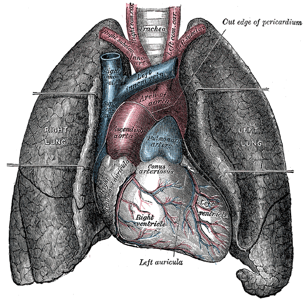
The heart sits in the middle mediastinum between the lungs, behind the sternum, above the diaphragm, and displaced slightly to the left of the midline. Its base points posterosuperiorly toward the right shoulder and its apex points anteroinferiorly, usually palpable in the fifth intercostal space at the midclavicular line. The fibrous pericardium anchors the organ and limits acute overdistension, while the serous pericardium folds into parietal and visceral layers separated by a thin film of lubricating fluid.
Four chambers handle two circulations in parallel. The right atrium receives deoxygenated blood from the superior vena cava, the inferior vena cava, and the coronary sinus. The right ventricle, with a wall of only 3 to 5 mm, ejects into the low-pressure pulmonary trunk. The left atrium receives oxygenated blood through four pulmonary veins, and the left ventricle, carrying 12 to 15 mm of dense myocardium, generates the pressure needed to perfuse the entire systemic circuit.
Four valves enforce one-way flow. The tricuspid and mitral atrioventricular valves are tethered by chordae tendineae to papillary muscles, which contract early in systole so the leaflets seal rather than prolapse. The pulmonary and aortic semilunar valves close passively when downstream pressure exceeds ventricular pressure. The myocardium itself is supplied by the left coronary artery, which divides into the left anterior descending and circumflex branches, and by the right coronary artery, which in most people also supplies the sinoatrial and atrioventricular nodes.
Physiology: how it works
Electrical activity originates in the sinoatrial node, where pacemaker cells depolarise spontaneously rather than waiting for an external signal. The impulse spreads across the atria, is deliberately delayed about 0.1 seconds at the atrioventricular node so atrial contraction can finish filling the ventricles, and is then distributed rapidly through the bundle of His, the bundle branches, and the Purkinje network. The fibrous skeleton insulates atria from ventricles electrically, making the atrioventricular bundle the only normal conduction path between them.
Contraction depends on calcium-induced calcium release. Depolarisation opens L-type calcium channels in the T-tubules; that trigger calcium opens ryanodine receptors on the sarcoplasmic reticulum, releasing a much larger internal store. Calcium binds troponin C, tropomyosin shifts off the actin binding sites, and ATP-powered actin-myosin cross-bridges cycle. Relaxation requires active removal of calcium by the SERCA2a pump and the sodium-calcium exchanger, which is why diastole consumes energy rather than being simple passive recoil.
At about 75 beats per minute a full cardiac cycle takes 0.8 seconds and passes through atrial systole, isovolumetric ventricular contraction, ejection, isovolumetric relaxation, and ventricular filling. Cardiac output equals heart rate multiplied by stroke volume, close to 5 litres per minute at rest. Stroke volume is set by preload, described by the Frank-Starling relationship, together with afterload and contractility, and each of those can be modulated independently by autonomic tone, circulating catecholamines, and drugs.
Biology: the living cells
Cardiomyocytes are short, branched, striated cells that usually carry a single central nucleus. Up to a third of their volume is mitochondria, because the myocardium is almost entirely aerobic and oxidises fatty acids, glucose, and lactate continuously. Their contractile machinery is built from actin, myosin, titin, troponin, and tropomyosin arranged into sarcomeres, with T-tubules carrying the surface membrane deep into the cell so calcium signalling stays synchronous. Intercalated discs join neighbouring cells through fascia adherens and desmosomes for mechanical continuity and connexin-43 gap junctions for electrical continuity, which lets the myocardium behave as a functional syncytium. Nodal cells trade contractile protein for automaticity, and Purkinje cells are large, glycogen-rich, and specialised for conduction speed rather than force.
Histology: under the microscope
The heart wall has three layers. The epicardium is a mesothelium over loose connective tissue carrying fat, coronary vessels, lymphatics, and autonomic nerves. The myocardium is the thick middle layer of cardiac muscle, arranged in helical sheets that let the ventricle wring rather than merely squeeze; its thickness varies dramatically from the thin atrial walls to the dense left ventricle. The endocardium is an endothelium continuous with the vascular lining, sitting on connective tissue and a subendocardial layer that carries vessels, nerves, and Purkinje fibres. Under the microscope the diagnostic features are branching striated fibres, central nuclei, and the dark transverse lines of intercalated discs, which together separate cardiac muscle from skeletal and smooth muscle.
Clinical correlations
Myocardial infarction follows occlusion of a coronary artery, usually by thrombus forming on a ruptured atherosclerotic plaque. Because cardiomyocytes have almost no regenerative capacity, ischaemic tissue is replaced by non-contractile fibrous scar and the functional loss is permanent. The territory affected is predictable from the vessel involved: the left anterior descending supplies the anterior wall and septum, the circumflex the lateral wall, and the right coronary artery the inferior wall and often the conduction nodes.
Heart failure describes a heart that cannot meet metabolic demand at normal filling pressures. It may reflect impaired contraction, seen as a reduced ejection fraction, or impaired filling with a preserved ejection fraction, and often both. The compensatory sympathetic and renin-angiotensin-aldosterone responses that support blood pressure acutely become maladaptive over months, driving remodelling, fibrosis, and progressive decline, which is why much of modern heart failure therapy works by blocking those same pathways.
Arrhythmias arise from abnormal impulse generation, abnormal conduction, or re-entry, and include atrial fibrillation, ventricular tachycardia, and the degrees of heart block. Valve disease loads the ventricle in two distinct ways: stenosis imposes a pressure load and drives concentric hypertrophy, while regurgitation imposes a volume load and drives eccentric dilatation. Across all of these, the baroreceptor reflex uses stretch receptors in the carotid sinus and aortic arch to adjust vagal and sympathetic outflow beat by beat.
Common questions about the heart
What are the four chambers of the heart and what does each one do?
The right atrium receives deoxygenated blood from the body, the right ventricle pumps it to the lungs, the left atrium receives oxygenated blood from the lungs, and the left ventricle pumps it to the rest of the body. The left ventricle has the thickest wall because it works against the highest pressure.
How does the heart generate its own heartbeat?
Pacemaker cells in the sinoatrial node depolarise spontaneously without any nerve input, setting the intrinsic rate. The signal reaches the atrioventricular node, which delays it briefly, then spreads through the His-Purkinje system to trigger a coordinated ventricular contraction. Nerves and hormones modulate this rate but do not create it.
What does cardiac muscle look like under a microscope?
Cardiac muscle shows short branching striated fibres with one or two central nuclei, separated by dark transverse intercalated discs. Those three features together distinguish it from skeletal muscle, which is unbranched with peripheral nuclei sitting at the edge of each fibre.
Why can the heart not repair itself after a heart attack?
Adult cardiomyocytes divide at an extremely low rate, so lost muscle is not meaningfully replaced. The damaged region heals with collagen scar instead, which provides structural strength but cannot contract or conduct, permanently reducing pump function in that territory.
Selected references
- Hall JE, Hall ME. Guyton and Hall Textbook of Medical Physiology. 14th ed. Elsevier; 2021.
- Standring S, ed. Gray's Anatomy: The Anatomical Basis of Clinical Practice. 42nd ed. Elsevier; 2021.
- Drake RL, Vogl AW, Mitchell AWM. Gray's Anatomy for Students. 4th ed. Elsevier; 2020.
- Bers DM. Cardiac excitation-contraction coupling. Nature. 2002;415(6868):198-205.
- Ross MH, Pawlina W. Histology: A Text and Atlas. 8th ed. Wolters Kluwer; 2020.
- Klabunde RE. Cardiovascular Physiology Concepts. 3rd ed. Wolters Kluwer; 2021.
Medical disclaimer. This article is written for education and general understanding. It is not medical advice and cannot replace assessment by a qualified healthcare professional. If you have symptoms or health concerns, speak with a clinician. Read the full disclaimer.