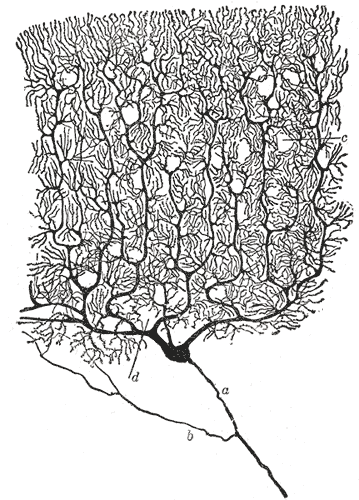
Nervous tissue looks like one thing and is really two. Neurons are the cells that carry information, and they get the attention: they generate electrical signals, connect to each other in networks of extraordinary complexity, and essentially never divide again after you are born. Glial cells are everything else, and there are at least as many of them. For a long time they were dismissed as packing — glia means glue — but they build the myelin that makes fast conduction possible, control the chemical environment neurons need, defend the tissue, and are the origin of most primary brain tumours. You cannot understand the first population without the second.
Where it is, and what it looks like
A neuron has three parts and they map onto three jobs. The cell body, or soma, holds the nucleus and almost all the synthetic machinery, and it is where the decision to fire is made. Dendrites are the branching processes that receive input, and their surface area is what allows a single neuron to collect signals from thousands of others. The axon is the single long output process, which may be a fraction of a millimetre or, in the case of the fibres running from the spinal cord to the foot, close to a metre. It ends in terminals that form synapses with the next cell. Neurons are classified by how many processes leave the soma: multipolar neurons, with one axon and many dendrites, make up most of the central nervous system; bipolar neurons serve the retina and olfactory epithelium; and pseudounipolar neurons, whose single process splits in two, carry sensation from the body into the spinal cord.
Nervous tissue is organised into grey matter and white matter, and the difference is myelin. Grey matter contains cell bodies, dendrites and synapses; it forms the cortex on the surface of the brain, the deep nuclei, and the butterfly-shaped core of the spinal cord. White matter contains the myelinated axons running between those regions and looks white to the naked eye because myelin is mostly lipid. The same pattern is inverted in the brain and the cord: grey is outside and white inside in the cerebrum, and the reverse in the spinal cord. Outside the central nervous system, cell bodies gather into ganglia and axons run in bundles as peripheral nerves.
Glial cells come in four types centrally and two peripherally. Astrocytes are the most numerous, star-shaped, and reach both the neurons and the capillaries. Oligodendrocytes make myelin in the central nervous system, each one wrapping segments of up to fifty different axons. Microglia are the resident immune cells, derived not from neural tissue but from the same lineage as macrophages. Ependymal cells line the ventricles and central canal, are ciliated, and produce and circulate cerebrospinal fluid. In the peripheral nervous system, Schwann cells myelinate axons — one cell to one internode of one axon, not fifty — and satellite cells surround the cell bodies in ganglia.
What it does
A resting neuron holds its inside at about minus 70 millivolts relative to the outside, maintained by the sodium-potassium ATPase pumping three sodium ions out for every two potassium ions in, and by the membrane being far more permeable to potassium than to sodium. That pump consumes more than half of a neuron's entire energy budget, which is why the brain uses roughly 20 per cent of the body's oxygen on 2 per cent of its mass, and why neurons die within minutes when blood flow stops.
An action potential is a brief, self-propagating reversal of that voltage. Depolarisation to threshold opens voltage-gated sodium channels, sodium rushes in and drives the membrane to around plus 30 millivolts, then those channels inactivate and voltage-gated potassium channels open, letting potassium out and restoring the resting state. The whole event takes about a millisecond, is all-or-nothing, and cannot be repeated during the refractory period that follows — which is what forces the signal to travel in one direction and sets a ceiling on firing rate. Information is carried in the frequency of these identical spikes, not their size.
Myelin makes this fast. A myelin sheath is an insulating wrap interrupted every millimetre or so by a bare patch, the node of Ranvier, where sodium channels are densely clustered. Current flows passively down the insulated internode and the action potential is regenerated only at the nodes, so the impulse jumps from node to node — saltatory conduction — reaching up to 120 metres per second in the largest myelinated fibres, against around one metre per second in unmyelinated ones of similar size. At the far end, the arriving action potential opens calcium channels in the terminal, vesicles of neurotransmitter fuse with the membrane and release their contents into the synaptic cleft, and receptors on the next cell convert the chemical signal back into an electrical one that is either excitatory or inhibitory. A neuron fires only when the sum of all such inputs reaches threshold at the axon hillock.
The cells that build it
The division of labour between neurons and glia is sharper than the old picture suggested. Astrocytes take up excess potassium and neurotransmitter from the extracellular space, so a synapse can be switched off cleanly and the next signal is not drowned in the last one; they supply neurons with lactate as a metabolic substrate; their end-feet wrap the capillaries and induce the tight junctions that create the blood-brain barrier; and after injury they proliferate to form the glial scar, which walls off damage but also blocks regrowing axons. Oligodendrocytes and Schwann cells both make myelin but differ in a way that matters clinically: an oligodendrocyte extends processes to many axons and cannot easily remyelinate after damage, while a Schwann cell serves a single internode and, together with the intact basal lamina tube it leaves behind, allows peripheral nerves to regenerate at roughly a millimetre a day. Microglia patrol continuously, prune unused synapses during development, and become the tissue's macrophages after injury. Neurons themselves are among the most specialised cells in the body and have paid for it: with a handful of exceptions, they leave the cell cycle permanently and cannot be replaced, so the response to a dead neuron is removal and scarring rather than replacement. That is also why primary tumours of the brain are almost never neuronal — gliomas arise from the cells that can still divide.
How to recognise it on a slide
In a stained section of grey matter, the neuronal cell bodies are the obvious feature: large, often triangular or pyramidal, with a strikingly pale round nucleus and a single dark nucleolus that stands out like an owl's eye. The cytoplasm contains coarse basophilic clumps, the Nissl substance, which is rough endoplasmic reticulum in quantities appropriate to a cell that must maintain a metre of axon. Nissl substance fills the soma and dendrites but stops abruptly at the axon hillock, which is a useful way to tell which process is the axon. Scattered densely between the neurons are far more numerous small dark nuclei with almost no visible cytoplasm — these are the glia, and on a routine haematoxylin and eosin section you can rarely tell one type from another without immunostaining. The pale pink felt-like background between all the cells is the neuropil, a dense mat of dendrites, axons and glial processes. White matter, by contrast, looks almost empty: rows of glial nuclei running between pale tracts, because the myelin lipid has been dissolved out during processing and leaves the axons as ghostly circles or lines depending on the plane of section.
When it goes wrong
Demyelinating disease shows how much depends on the insulation rather than the wire. In multiple sclerosis, immune attack destroys central myelin in discrete plaques scattered through the brain, optic nerves and spinal cord, and because oligodendrocytes remyelinate poorly the deficits accumulate. Symptoms are separated in time and in place, which is exactly what you would predict from lesions appearing at different sites on different occasions. Guillain-Barré syndrome is the peripheral counterpart, usually triggered by a preceding infection, and it produces an ascending weakness that starts in the legs — but because Schwann cells remyelinate well, most patients recover substantially.
Because neurons cannot be replaced, any event that kills them leaves permanent loss. In ischaemic stroke, neurons in the core die within minutes and are visible on a slide as red neurons within six to twelve hours, after which the brain undergoes liquefactive necrosis and leaves a fluid-filled cavity walled off by reactive astrocytes. In the neurodegenerative diseases the same endpoint is reached slowly and selectively: dopaminergic neurons of the substantia nigra in Parkinson's disease, cortical and hippocampal neurons in Alzheimer's disease, motor neurons in amyotrophic lateral sclerosis. In each, function is lost in the pattern dictated by which population dies.
The glial cells that retain the ability to divide are the ones that become tumours. Gliomas — astrocytomas, of which glioblastoma is the most aggressive, along with oligodendrogliomas and ependymomas — make up the large majority of primary brain tumours in adults, while true neuronal tumours are rare. Glia also determine what medicine can reach the brain at all: the blood-brain barrier, built on tight junctions induced by astrocyte end-feet, excludes most large and water-soluble drugs, which is a central problem in treating both brain infection and brain cancer, and is why some antibiotics simply cannot be used for meningitis.
Common questions about nervous tissue
What is the difference between neurons and glial cells?
Neurons generate and transmit electrical signals and form the circuits that process information; they are highly specialised and, with rare exceptions, cannot divide or be replaced. Glial cells support them: astrocytes manage the chemical environment and build the blood-brain barrier, oligodendrocytes and Schwann cells make myelin, microglia act as the immune cells, and ependymal cells produce and circulate cerebrospinal fluid. Glia retain the ability to divide, which is why they are the source of most brain tumours.
What are the four types of glial cell in the central nervous system?
Astrocytes, which regulate the extracellular environment, supply neurons metabolically, and induce the blood-brain barrier; oligodendrocytes, which produce central myelin; microglia, the resident immune cells, derived from the macrophage lineage rather than from neural tissue; and ependymal cells, which line the ventricles and central canal and circulate cerebrospinal fluid. In the peripheral nervous system the equivalents are Schwann cells and satellite cells.
Why do peripheral nerves regenerate but central ones do not?
Schwann cells survive the injury, clear debris, and leave behind an intact basal lamina tube that guides a regrowing axon back toward its target at roughly a millimetre a day. In the central nervous system, oligodendrocytes remyelinate poorly, myelin debris contains molecules that actively inhibit axon growth, and reactive astrocytes form a glial scar that walls the lesion off. The neuron's own capacity to regrow is similar; the environment is what differs.
What is the difference between grey matter and white matter?
Grey matter contains neuronal cell bodies, dendrites and synapses — it is where processing happens. White matter contains the myelinated axons that carry signals between regions, and it looks white because myelin is largely lipid. In the cerebrum, grey matter forms the outer cortex with white matter inside; in the spinal cord the arrangement is reversed, with grey matter in a central butterfly shape surrounded by white.
Selected references
- Ross MH, Pawlina W. Histology: A Text and Atlas. 8th ed. Wolters Kluwer; 2020.
- Kandel ER, Koester JD, Mack SH, Siegelbaum SA. Principles of Neural Science. 6th ed. McGraw Hill; 2021.
- Standring S, ed. Gray's Anatomy: The Anatomical Basis of Clinical Practice. 42nd ed. Elsevier; 2021.
- von Bartheld CS, Bahney J, Herculano-Houzel S. The search for true numbers of neurons and glial cells in the human brain. Journal of Comparative Neurology. 2016;524(18):3865-3895.
- Abbott NJ, Patabendige AA, Dolman DE, Yurdakul SR, Begley DJ. Structure and function of the blood-brain barrier. Neurobiology of Disease. 2010;37(1):13-25.
- Kumar V, Abbas AK, Aster JC. Robbins and Cotran Pathologic Basis of Disease. 10th ed. Elsevier; 2021.
Medical disclaimer. This article is written for education and general understanding. It is not medical advice and cannot replace assessment by a qualified healthcare professional. If you have symptoms or health concerns, speak with a clinician. Read the full disclaimer.