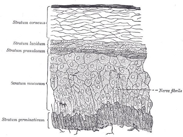
Epithelium is the tissue that draws the line between inside and outside. It covers every external surface, lines every internal cavity and tube, and forms every gland in the body, which means that anything entering or leaving you has to cross an epithelial cell to do it. Almost all of that work is done by sheets of cells packed so tightly that there is no room for a blood vessel between them. The naming looks intimidating until you notice that it is only ever answering two questions: how many layers are there, and what shape are the cells at the free surface?
Where it is, and what it looks like
Epithelia fall into three jobs. Covering and lining epithelium forms the outer layer of skin, the lining of the gut, airways, blood vessels and body cavities, and the surface of organs. Glandular epithelium is what you get when a covering epithelium grows down into the tissue beneath and specialises in secretion — exocrine glands keep a duct to the surface, endocrine glands lose theirs and release into the blood instead. Sensory epithelium, in the nose, tongue, inner ear and retina, is covering epithelium that has taken on receptor cells. All three sit on a basement membrane and none of them contain blood vessels.
The first half of every epithelium's name counts the layers. Simple means one layer, with every cell touching the basement membrane, and it is what you find wherever material has to move across quickly. Stratified means two or more layers, and it appears wherever the surface takes physical punishment; only the deepest layer touches the basement membrane, and the type is named for the shape of the cells at the top, not the bottom. Pseudostratified looks layered but is not: every cell reaches the basement membrane, but they are different heights, so their nuclei sit at different levels and give a false impression of stacking.
The second half names the shape of the surface cells. Squamous cells are flat and wide, like floor tiles seen edge-on. Cuboidal cells are roughly as tall as they are wide, with a round central nucleus. Columnar cells are taller than they are wide, usually with an oval nucleus near the base. Put the two halves together and the map falls out: simple squamous in the alveoli, capillaries and body cavities; simple cuboidal in kidney tubules and small gland ducts; simple columnar through the stomach and intestines; pseudostratified ciliated columnar in the trachea and bronchi; stratified squamous in the skin, mouth, oesophagus, anus and vagina; and transitional, or urothelium, in the renal pelvis, ureters and bladder, where the cells change shape as the organ fills.
What it does
The first function is barrier. A continuous sheet of cells sealed to its neighbours decides what crosses and what does not, and the seal is not metaphorical: tight junctions form a belt near the apical surface of every epithelial cell and physically occlude the space between cells, so anything crossing the sheet must pass through a cell rather than around it. That is what allows an epithelium to keep a hostile lumen — stomach acid, urine, faecal bacteria, the outside world — a few micrometres away from living tissue, and it is why the skin's stratified squamous epithelium is many layers thick while the alveolar lining is a single flattened cell.
The second is selective transport, and it depends on polarity. An epithelial cell is not symmetrical: its apical membrane, facing the lumen, carries a different set of pumps, channels and enzymes from its basolateral membrane, facing the blood. Because tight junctions physically stop membrane proteins drifting from one domain to the other, the cell can take a substance in at the top and release it at the bottom, moving material in a single direction across the whole sheet. That one arrangement produces glucose absorption in the intestine, sodium reabsorption in the kidney, and acid secretion in the stomach.
The third is secretion, and the fourth is movement. Glandular epithelium manufactures and exports mucus, enzymes, hormones, sweat, milk and bile. Ciliated epithelium sweeps material along a surface: several hundred cilia on each cell of the airway lining beat in coordinated waves at roughly ten to twenty times a second, carrying a sheet of mucus and trapped debris up towards the throat, and the same mechanism moves the ovum along the uterine tube. Epithelium also carries most of the body's sensory receptors, and, in the gut and airways, a population of hormone-releasing cells that report on what is passing through.
The cells that build it
Epithelial cells are held together and held down by a specific set of junctions, and knowing them explains most epithelial disease. Tight junctions seal the space between neighbouring cells and separate apical from basolateral membrane. Adherens junctions just below them link the actin cytoskeletons of adjacent cells through cadherins, and desmosomes do the same for intermediate filaments, spot-welding cells so a sheet can take shear without tearing. Gap junctions let small molecules and ions pass directly between cells, coupling them chemically and electrically. Hemidesmosomes and focal adhesions anchor the base of the cell to the basement membrane, a sheet of type IV collagen, laminin and proteoglycans that the epithelium and the underlying connective tissue build together. Because no capillary crosses that membrane, every epithelial cell is fed by diffusion from vessels in the connective tissue beneath, which puts a hard ceiling on how thick an epithelium can be. Epithelium is also the fastest-renewing tissue in the body: the intestinal lining is completely replaced every three to five days from stem cells at the base of the crypts, and the epidermis turns over roughly monthly. That turnover is what makes epithelium so good at repair and so prone to cancer.
How to recognise it on a slide
On a slide, epithelium is the tissue where the cells are crowded and the space between them is not. Find a free surface — a lumen, an outer edge — and follow the cells down to a thin line where they stop abruptly: that line is the basement membrane, and everything above it is epithelium. There are no capillaries inside the sheet, which is the quickest way to separate epithelium from the connective tissue below, where nuclei are sparse and vessels are obvious. Then read the name off the picture. Count the layers first: if every nucleus sits at the same level on the basement membrane it is simple, and if nuclei are stacked at different heights you must decide between stratified and pseudostratified — look for cilia and goblet cells, which point to pseudostratified, and for a flattened surface layer, which points to stratified squamous. Then judge the shape of the topmost cells only. Flattened nuclei parallel to the surface mean squamous, round central nuclei mean cuboidal, and tall oval nuclei lined up near the base mean columnar. Transitional epithelium gives itself away by large rounded dome-shaped surface cells, some of them binucleate, that flatten out when the organ is distended.
When it goes wrong
Because epithelium divides so often, it is where most cancers begin: carcinomas, which arise from epithelium, account for roughly nine in ten human cancers, and the common ones — lung, breast, colon, prostate, skin — are all epithelial. The sequence is usually visible under the microscope long before it becomes dangerous, running from hyperplasia through dysplasia, in which cells lose their orderly layering and uniform appearance, to carcinoma in situ, which is still confined above the basement membrane, and finally to invasive carcinoma once that membrane is breached. Every screening programme in medicine — cervical smears, colonoscopy, mammography — exists to catch that sequence before the last step.
Epithelium can also change type. Metaplasia is the replacement of one mature epithelium by another that is better suited to a new environment, and it is adaptive rather than malignant. Chronic acid reflux converts the stratified squamous lining of the lower oesophagus to intestinal-type columnar epithelium, which is Barrett's oesophagus; cigarette smoke converts the pseudostratified ciliated epithelium of the bronchi to stratified squamous, which resists smoke but can no longer clear mucus. Both are reversible in principle, and both carry a raised long-term cancer risk, which is why they are followed rather than ignored.
Damage to the epithelial barrier itself produces a recognisable set of illnesses. In coeliac disease an immune response to gluten flattens the intestinal villi and destroys the absorptive surface, causing malabsorption out of proportion to the length of gut involved. In cystic fibrosis a faulty chloride channel in epithelial cell membranes leaves airway and pancreatic secretions thick and immovable. In severe burns, loss of the epidermis produces fluid loss and infection rather than pain as the immediate threat. And in the gut, breakdown of tight junctions lets luminal bacteria and their products reach the bloodstream, one of the routes by which an intestinal problem becomes a systemic one.
Common questions about epithelial tissue
What are the main types of epithelial tissue?
Epithelia are classified by layers and by the shape of their surface cells. Simple squamous lines alveoli, capillaries and body cavities; simple cuboidal lines kidney tubules and gland ducts; simple columnar lines the stomach and intestines; pseudostratified ciliated columnar lines the trachea and bronchi; stratified squamous covers the skin, mouth, oesophagus and vagina; and transitional epithelium, or urothelium, lines the renal pelvis, ureters and bladder.
How do you identify epithelium under a microscope?
Look for a free surface, densely packed cells with very little material between them, and a sharp basement membrane where the sheet stops. Epithelium contains no blood vessels, so if you can see capillaries among the cells you are looking at connective tissue instead. Once you have found the sheet, count the layers, then judge the shape of the cells at the surface only.
What is the difference between stratified and pseudostratified epithelium?
In stratified epithelium the cells really are stacked, and only the deepest layer touches the basement membrane. In pseudostratified epithelium every cell reaches the basement membrane, but the cells are different heights, so their nuclei sit at different levels and create the illusion of layering. Cilia and goblet cells are a strong clue for pseudostratified, since it is the classic airway lining.
Why does epithelium have no blood supply?
Blood vessels would breach the seal the tissue exists to maintain. Instead the epithelium sits on a basement membrane, and oxygen and nutrients diffuse across it from capillaries in the connective tissue beneath. The trade-off is a strict limit on thickness, which is why gas-exchange surfaces are a single flattened cell thick and why thick epithelia like the epidermis have a dead, non-metabolising surface layer.
Selected references
- Ross MH, Pawlina W. Histology: A Text and Atlas. 8th ed. Wolters Kluwer; 2020.
- Mescher AL. Junqueira's Basic Histology: Text and Atlas. 16th ed. McGraw Hill; 2021.
- Young B, O'Dowd G, Woodford P. Wheater's Functional Histology. 6th ed. Elsevier; 2014.
- Alberts B, Heald R, Johnson A, et al. Molecular Biology of the Cell. 7th ed. W. W. Norton; 2022.
- Kumar V, Abbas AK, Aster JC. Robbins and Cotran Pathologic Basis of Disease. 10th ed. Elsevier; 2021.
- Zihni C, Mills C, Matter K, Balda MS. Tight junctions: from simple barriers to multifunctional molecular gates. Nature Reviews Molecular Cell Biology. 2016;17(9):564-580.
Medical disclaimer. This article is written for education and general understanding. It is not medical advice and cannot replace assessment by a qualified healthcare professional. If you have symptoms or health concerns, speak with a clinician. Read the full disclaimer.