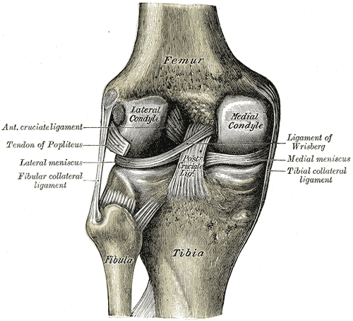
Bone looks inert and behaves as though it were, but it is one of the most dynamic tissues in the body. It supports and protects, it levers the body into motion, it houses the marrow that makes blood cells, and it serves as the reservoir holding roughly 99 percent of the body's calcium. Around 10 percent of the adult skeleton is replaced every year, and the shape it takes is continuously negotiated with the mechanical loads placed on it.
Anatomy: the structure
Bones are classified by shape into long, short, flat, irregular, and sesamoid. A long bone has a diaphysis, or shaft, with a central medullary cavity, expanded epiphyses at each end covered by articular cartilage, and a metaphysis between them containing the epiphyseal growth plate during growth and its residual line afterwards. The outer surface is covered by periosteum, a fibrous outer layer with an inner osteogenic layer anchored by Sharpey fibres, while the inner surfaces are lined by endosteum. Periosteum is richly innervated, which is why periosteal injury is disproportionately painful.
At the tissue level, bone exists in two architectural forms. Compact or cortical bone forms the dense outer shell and about 80 percent of skeletal mass, optimised for resisting bending and torsion. Trabecular, cancellous, or spongy bone forms a three-dimensional lattice inside the epiphyses and within flat bones, with far greater surface area for metabolic exchange and trabeculae aligned along lines of principal stress. Trabecular bone has a much higher turnover rate, which is why osteoporotic fractures occur first at trabecular-rich sites such as the vertebral bodies, femoral neck, and distal radius.
Bone is a composite material, and its two components divide the work. The organic matrix, about 35 percent by weight and roughly 90 percent type I collagen with proteoglycans and non-collagenous proteins such as osteocalcin and osteopontin, supplies tensile strength and flexibility. The inorganic phase, about 65 percent, consists of hydroxyapatite crystals of calcium and phosphate that supply compressive strength and hardness. Removing the mineral leaves a bone that bends like rubber; removing the collagen leaves one that shatters like chalk. Blood arrives through nutrient arteries entering the diaphysis, metaphyseal and epiphyseal vessels, and periosteal vessels supplying the outer cortex.
Physiology: how it works
Bone forms in two ways during development. Intramembranous ossification builds flat bones of the skull, the mandible, and the clavicle directly from mesenchymal condensations without a cartilage stage. Endochondral ossification builds most of the rest of the skeleton on a hyaline cartilage model, with a primary ossification centre in the diaphysis and secondary centres in the epiphyses. Longitudinal growth then occurs at the epiphyseal plate through an orderly sequence of zones: resting, proliferation, hypertrophy, calcification, and ossification. Growth stops when the plate closes under the influence of sex hormones, while appositional growth in width continues from the periosteum throughout life.
The skeleton is the body's calcium bank, and three hormones control the balance. Parathyroid hormone, released when plasma calcium falls, indirectly stimulates osteoclastic resorption, increases renal calcium reabsorption, and promotes activation of vitamin D. Calcitriol, the active form of vitamin D, increases intestinal calcium and phosphate absorption, which is its dominant effect. Calcitonin from thyroid C cells inhibits osteoclasts when calcium is high, though its role in adult humans is limited. Because plasma calcium must be held within a narrow range for nerve and muscle function, the skeleton will be sacrificed to defend it if dietary intake is inadequate.
Remodelling is continuous and organised. Basic multicellular units work in a coupled sequence of activation, resorption by osteoclasts over about three weeks, reversal, and formation by osteoblasts over about three months, so formation is always the slower half of the cycle. Osteocytes embedded in the matrix act as the mechanosensors that direct where this happens, detecting fluid flow through the canaliculi during loading and adjusting signalling accordingly. This is the cellular basis of Wolff law: loaded bone is reinforced, and unloaded bone, whether from bed rest, immobilisation, or spaceflight, is resorbed. Marrow within the medullary cavity contributes the separate function of haematopoiesis, with red marrow progressively replaced by yellow fatty marrow with age except in the axial skeleton and proximal femur.
Biology: the living cells
Four cell types build and maintain bone. Osteoprogenitor cells in the periosteum, endosteum, and marrow stroma are mesenchymal stem cells that supply new osteoblasts. Osteoblasts are cuboidal cells lining bone surfaces that secrete osteoid, the unmineralised organic matrix, then control its mineralisation, and they carry receptors for parathyroid hormone and vitamin D. Osteocytes are osteoblasts that have become trapped within the matrix they made; they occupy small spaces called lacunae, extend processes through channels called canaliculi to contact neighbours through gap junctions, and make up over 90 percent of bone cells while sensing mechanical strain and regulating local remodelling. Osteoclasts are large multinucleated cells derived from the monocyte-macrophage lineage rather than from mesenchyme; they attach to bone through a sealing zone, develop a ruffled border, and resorb matrix by secreting hydrogen ions to dissolve mineral and cathepsin K to digest collagen. The coupling between them runs through RANK ligand expressed by osteoblasts, which activates osteoclast precursors, and osteoprotegerin, a decoy receptor that blocks it, and this axis is the target of modern antiresorptive therapy.
Histology: under the microscope
Mature bone is lamellar, meaning the collagen is laid down in parallel sheets with alternating fibre orientation, which resists force from multiple directions. In compact bone the fundamental unit is the osteon, or Haversian system: concentric lamellae arranged around a central Haversian canal carrying vessels and nerves, with osteocytes in lacunae between lamellae and canaliculi radiating outward like spokes. Volkmann canals run transversely, connecting Haversian canals to each other and to the periosteal and endosteal surfaces. Interstitial lamellae, the remnants of osteons removed by previous remodelling cycles, fill the spaces between intact osteons and are a visible record of turnover, while circumferential lamellae wrap the outer and inner surfaces of the whole shaft. Trabecular bone contains no osteons because the trabeculae are thin enough to be nourished by diffusion from marrow; it is built from parallel lamellae instead. Woven bone, by contrast, has randomly arranged collagen and is mechanically weaker; it is normal in the fetus and in fracture callus but abnormal in adult tissue, where it indicates rapid pathological formation.
Clinical correlations
Osteoporosis is a quantitative disorder: bone mass falls and microarchitecture deteriorates while the remaining bone is normally mineralised. It results from an imbalance in which resorption outpaces formation, accelerated by oestrogen withdrawal after menopause, by age, and by glucocorticoid use. Because trabecular bone turns over faster, fractures appear first at the vertebrae, femoral neck, and distal radius. Osteomalacia, and rickets in children, is a qualitative disorder instead: vitamin D deficiency leaves osteoid inadequately mineralised, producing soft bone that deforms rather than simply breaking.
Fracture healing recapitulates development in four overlapping stages. A haematoma forms and triggers inflammation; a soft callus of fibrocartilage bridges the gap; endochondral ossification converts it into a hard callus of woven bone; and remodelling gradually replaces woven bone with lamellar bone aligned to load, a process that can continue for years. Healing depends on blood supply and mechanical stability, which is why displaced fractures of the scaphoid and femoral neck, where retrograde blood supply is easily interrupted, risk avascular necrosis and non-union.
Endocrine and neoplastic disease also write themselves into bone. Primary hyperparathyroidism drives excessive resorption with subperiosteal erosions and, in advanced cases, brown tumours. Paget disease produces disordered accelerated turnover with a characteristic mosaic pattern of cement lines on histology. Metastatic disease reaches the axial skeleton preferentially, since red marrow is where blood-borne cells lodge, and produces lytic lesions from most primaries but characteristically sclerotic lesions from prostate carcinoma.
Common questions about bone
Is bone a living tissue?
Yes. Bone contains living cells throughout its matrix, has its own blood supply and sensory nerves, remodels continuously in response to mechanical load and hormonal signals, and can repair itself after fracture. Roughly a tenth of the adult skeleton is replaced every year.
What is an osteon?
An osteon, or Haversian system, is the structural unit of compact bone. It consists of concentric rings of lamellar bone around a central canal carrying blood vessels and nerves, with osteocytes sitting in lacunae between the rings and communicating through fine canaliculi.
What is the difference between osteoporosis and osteomalacia?
Osteoporosis means there is less bone, but what remains is normally mineralised, so the problem is quantity. Osteomalacia means there is a normal amount of matrix that is inadequately mineralised, usually through vitamin D deficiency, so the problem is quality and the bone becomes soft rather than brittle.
How do osteoblasts and osteoclasts work together?
They operate as a coupled unit. Osteoclasts resorb a packet of old bone over about three weeks, then osteoblasts fill the cavity with new matrix over about three months. Osteoblasts control the process by expressing RANK ligand, which activates osteoclasts, and osteoprotegerin, which blocks that activation.
Selected references
- Standring S, ed. Gray's Anatomy: The Anatomical Basis of Clinical Practice. 42nd ed. Elsevier; 2021.
- Ross MH, Pawlina W. Histology: A Text and Atlas. 8th ed. Wolters Kluwer; 2020.
- Hall JE, Hall ME. Guyton and Hall Textbook of Medical Physiology. 14th ed. Elsevier; 2021.
- Boyle WJ, Simonet WS, Lacey DL. Osteoclast differentiation and activation. Nature. 2003;423(6937):337-342.
- Bonewald LF. The amazing osteocyte. Journal of Bone and Mineral Research. 2011;26(2):229-238.
- Kumar V, Abbas AK, Aster JC. Robbins and Cotran Pathologic Basis of Disease. 10th ed. Elsevier; 2021.
Medical disclaimer. This article is written for education and general understanding. It is not medical advice and cannot replace assessment by a qualified healthcare professional. If you have symptoms or health concerns, speak with a clinician. Read the full disclaimer.