The definition most people carry is that arteries carry oxygenated blood and veins carry deoxygenated blood, and it is wrong twice over: the pulmonary artery carries deoxygenated blood to the lungs, and the pulmonary veins carry oxygenated blood back. The real definition is about direction. Arteries carry blood away from the heart and veins carry it back, whatever is dissolved in it. Everything else that distinguishes them — wall thickness, lumen size, valves, how much blood each holds — follows from one fact: arteries work at high pressure and veins do not.
Where it is, and what it looks like
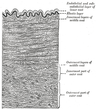
Every vessel larger than a capillary has the same three coats, and the differences between vessel types are differences in the proportions. The tunica intima is the innermost layer: a single sheet of endothelium on a thin basement membrane and a little subendothelial connective tissue, bounded in arteries by an internal elastic lamina. The tunica media is the middle layer of circular smooth muscle and elastic tissue, and it is the layer that varies most. The tunica adventitia, or externa, is the outer coat of connective tissue that anchors the vessel to its surroundings; in vessels above about a millimetre across it carries its own small blood supply, the vasa vasorum, because the wall is too thick to be fed by diffusion from the lumen.
Arteries come in three sizes with different priorities. Elastic, or conducting, arteries — the aorta and its largest branches — have a media dominated by sheets of elastin, which lets them balloon during systole and recoil during diastole, converting the heart's intermittent output into continuous flow. Muscular, or distributing, arteries such as the radial, femoral and coronary arteries have a media dominated by smooth muscle and route blood to specific organs. Arterioles, the smallest, have only one to three layers of smooth muscle but are the principal resistance vessels: because resistance varies with the fourth power of radius, a small change in arteriolar diameter has an enormous effect, and arterioles are therefore where blood pressure is actually set.
Veins run the same sequence in reverse and are built for capacity rather than pressure. Venules collect from capillaries, and the smallest of them are the site where white cells leave the circulation during inflammation. Small and medium veins have a thin media and a comparatively thick adventitia, and many of them — particularly in the limbs — contain valves, paired semilunar folds of intima that prevent backflow and make it possible for muscle contraction to push blood upward against gravity. Large veins such as the venae cavae have a well-developed adventitia containing longitudinal smooth muscle. At any moment roughly 60 to 70 per cent of the body's blood is sitting in the venous system, which is why veins are described as capacitance vessels.
What it does
The pressure difference explains almost every structural difference. Mean arterial pressure is around 90 to 100 millimetres of mercury and swings between systolic and diastolic with every beat; venous pressure is under 10 and barely pulsates. An artery therefore needs a thick muscular wall to contain that pressure without bursting, and it holds its round shape even when empty, which is why an artery in a histology section looks circular with a small lumen while the vein beside it looks flattened and irregular with a large one. The elastic recoil of the great arteries during diastole is also what keeps coronary perfusion going, since the coronary arteries fill between beats rather than during them.
Because venous pressure is so low, veins need help. Three mechanisms return blood to the heart against gravity. The skeletal muscle pump squeezes deep veins during contraction and, because the valves permit flow in one direction only, each squeeze moves a bolus toward the heart. The respiratory pump lowers intrathoracic pressure during inspiration, drawing blood into the chest. And venous tone, controlled by sympathetic nerves acting on the thin smooth muscle layer, can constrict the venous reservoir to shift blood back into the circulation — which is how the body compensates for the first litre of blood loss without any fall in blood pressure at all.
The capillaries between them are where the entire cardiovascular system justifies itself. A capillary is a single layer of endothelium on a basement membrane, five to ten micrometres across, so a red cell must deform to pass through. There are around ten billion of them with a combined surface area of several hundred square metres, and because that vast cross-sectional area slows flow to under a millimetre per second, blood spends one to three seconds in a capillary — long enough for exchange. Fluid movement across the wall follows Starling's principle: hydrostatic pressure pushes fluid out at the arterial end, colloid osmotic pressure from plasma proteins pulls most of it back at the venous end, and the small surplus is collected by lymphatics. Disturb any term in that balance and you get oedema.
The cells that build it
The endothelium is the one component every vessel shares, and it is far more than a lining. A continuous sheet of endothelial cells weighing about a kilogram in total, it secretes nitric oxide and prostacyclin to relax the underlying smooth muscle and endothelin to constrict it, so local blood flow is largely set by the vessel's own lining responding to shear stress and to signals from the tissue around it. It presents an anticoagulant surface in health, carrying heparan sulphate and thrombomodulin, and switches to a procoagulant, adhesive surface when injured or inflamed, expressing selectins and integrin ligands that let white cells roll, stick and cross into the tissue. Capillaries come in three structural forms matched to their organ: continuous capillaries, with unbroken endothelium and tight junctions, in muscle, lung, skin and, at their tightest, the brain, where they form the blood-brain barrier; fenestrated capillaries, perforated by small pores, in the kidney glomerulus, intestinal villi and endocrine glands, where rapid bulk exchange is needed; and sinusoidal capillaries, wide and leaky with gaps between cells and an incomplete basement membrane, in the liver, spleen and bone marrow, where whole cells and large proteins must pass. Around the capillaries sit pericytes, contractile cells sharing the basement membrane, which regulate flow and can differentiate into new vessel-wall cells during angiogenesis.
How to recognise it on a slide
Arteries and veins almost always run together, and the easiest way to tell them apart on a slide is to compare the two in the same field. The artery is the one that is round, with a thick wall and a relatively small lumen that often appears empty; the vein is flattened or collapsed, with a thin wall and a wide lumen usually full of red cells. Look next at the layers. In a muscular artery the tunica media is thick and obviously muscular, and it is bounded on its inner side by a wavy, refractile internal elastic lamina that is one of the most distinctive structures in histology — the crinkled appearance is a fixation artefact caused by the muscle contracting after death, and it is diagnostic. Veins have a thin media, no prominent internal elastic lamina, and an adventitia that is often thicker than the media, so the outer connective tissue dominates the wall. In an elastic artery such as the aorta the media is filled with parallel wavy sheets of elastin, best shown with an elastic stain, rather than with obvious layers of muscle. Capillaries are recognised by having no wall at all beyond a single endothelial nucleus bulging into a lumen just wide enough for one red cell.
When it goes wrong
Atherosclerosis is a disease of arteries and, specifically, of the intima. Endothelial injury from hypertension, smoking, diabetes or high LDL cholesterol allows lipid into the subendothelial space, where macrophages engulf it and become foam cells; smooth muscle migrates in from the media and lays down a fibrous cap over the resulting lipid core. That plaque narrows the lumen gradually, but the dangerous event is sudden: rupture of the cap exposes highly thrombogenic material, a clot forms within minutes, and the artery occludes. The same process in different territories produces myocardial infarction, ischaemic stroke and peripheral arterial disease. An aneurysm is the opposite failure, in which the media weakens and the wall dilates until it tears.
Venous disease is a failure of the return mechanisms rather than of the wall. Incompetent valves in the superficial leg veins allow blood to fall back down, raising pressure in the veins below and producing varicose veins, ankle swelling, skin discoloration and eventually venous ulceration. Deep vein thrombosis follows Virchow's triad — stasis, endothelial injury and hypercoagulability — which is why immobility, surgery, pregnancy and malignancy are the classic risk factors, and its danger is that a fragment can travel through the right heart and lodge in the pulmonary arteries as a pulmonary embolism.
Capillary problems present as fluid in the wrong place. Oedema results whenever the Starling balance is disturbed: raised venous pressure in heart failure, low plasma albumin in liver disease or nephrotic syndrome, increased permeability in inflammation and sepsis, or blocked lymphatic drainage after node clearance or in filariasis. And angiogenesis — the growth of new capillaries — is the process solid tumours must switch on to grow beyond a couple of millimetres, which is why drugs targeting vascular endothelial growth factor are used in cancer, and in the eye in wet macular degeneration and diabetic retinopathy.
Common questions about the blood vessels
What is the difference between arteries and veins?
Arteries carry blood away from the heart and veins carry it back, regardless of oxygen content. Arteries work at high pressure, so they have thick muscular walls, narrow round lumens and no valves. Veins work at low pressure, so they have thin walls, wide lumens, valves in the limbs to prevent backflow, and they hold most of the body's blood. On a slide the artery is round with a thick wall; the vein beside it is flattened with a thin one.
Do all arteries carry oxygenated blood?
No. The pulmonary arteries carry deoxygenated blood from the right ventricle to the lungs, and the umbilical arteries in a fetus carry deoxygenated blood to the placenta. Conversely the pulmonary veins and the umbilical vein carry oxygenated blood. Direction of flow relative to the heart is the definition; oxygen content is only the usual case.
What are the three layers of a blood vessel?
The tunica intima, an endothelial lining on a basement membrane with a little connective tissue; the tunica media, a middle layer of circular smooth muscle and elastic tissue; and the tunica adventitia, an outer connective tissue coat anchoring the vessel to its surroundings. Arteries have a thick media, veins a thin media and a relatively thick adventitia, and capillaries have only the endothelium and its basement membrane.
What do capillaries do?
All the actual exchange. A capillary wall is a single endothelial cell layer, thin enough for oxygen, carbon dioxide, glucose, ions and water to cross. Their combined cross-sectional area is so large that blood slows to under a millimetre per second and spends one to three seconds in each capillary, which is the time exchange needs. Continuous capillaries are the tightest, fenestrated ones are perforated for bulk filtration in the kidney and gut, and sinusoidal ones in the liver, spleen and marrow are leaky enough to let whole cells through.
Selected references
- Ross MH, Pawlina W. Histology: A Text and Atlas. 8th ed. Wolters Kluwer; 2020.
- Hall JE, Hall ME. Guyton and Hall Textbook of Medical Physiology. 14th ed. Elsevier; 2021.
- Standring S, ed. Gray's Anatomy: The Anatomical Basis of Clinical Practice. 42nd ed. Elsevier; 2021.
- Pober JS, Sessa WC. Evolving functions of endothelial cells in inflammation. Nature Reviews Immunology. 2007;7(10):803-815.
- Levick JR, Michel CC. Microvascular fluid exchange and the revised Starling principle. Cardiovascular Research. 2010;87(2):198-210.
- Kumar V, Abbas AK, Aster JC. Robbins and Cotran Pathologic Basis of Disease. 10th ed. Elsevier; 2021.
Medical disclaimer. This article is written for education and general understanding. It is not medical advice and cannot replace assessment by a qualified healthcare professional. If you have symptoms or health concerns, speak with a clinician. Read the full disclaimer.