Every other system in the body keeps the individual alive. This one does nothing for the individual at all — you can live a full life without it — and exists entirely for the generation after. That difference shows in how it is built. It stays dormant for the first decade or more of life, is switched on by a hormonal cascade at puberty, runs on cycles rather than steady states, and in one sex shuts down again in mid-life. It is also the only system whose anatomy differs fundamentally between sexes, though both versions develop from the same embryonic structures and are organised around the same three jobs: make gametes, make hormones, and bring the two gametes together.
The parts, and how they fit together
The male system is arranged around the fact that sperm production needs a temperature two to three degrees below core body temperature, which is why the testes sit outside the abdomen in the scrotum. Each testis contains around 800 tightly coiled seminiferous tubules where sperm are made, with interstitial Leydig cells between them producing testosterone. Sperm pass into the epididymis, a six-metre coiled tube on the back of the testis where they mature and gain motility over about two weeks, then through the vas deferens, which loops up into the pelvis. Three accessory glands supply the fluid: the seminal vesicles contribute around 60 per cent, rich in fructose to fuel the sperm; the prostate adds roughly 30 per cent, alkaline to neutralise vaginal acidity; and the bulbourethral glands add lubricant. Sperm themselves are only a small fraction of the volume.
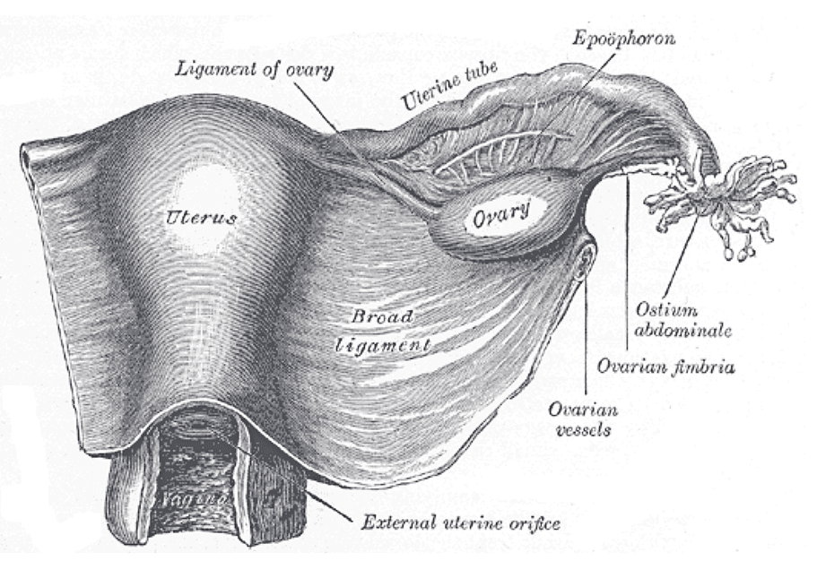
The female system is arranged around supporting a pregnancy rather than producing gametes continuously. The two ovaries produce oocytes and the hormones oestrogen and progesterone. The uterine tubes are not attached to the ovaries but open near them, their fimbriae sweeping the released oocyte inward; fertilisation normally happens in the ampulla, the widest part. The uterus is a thick-walled muscular organ with three layers — an outer perimetrium, a thick myometrium of smooth muscle, and an endometrium that is rebuilt and shed each cycle. Its lower narrow part, the cervix, projects into the vagina and produces mucus whose consistency changes across the cycle. The vagina is a muscular canal that serves as the birth canal and receives sperm.
Both systems arise from the same starting material, which explains a set of structural parallels that are otherwise puzzling. The indifferent gonad becomes testis or ovary depending on whether the SRY gene on the Y chromosome is present; the testis then secretes testosterone and anti-Müllerian hormone, and it is the presence or absence of those signals rather than the chromosomes directly that determines which duct system survives. The Wolffian ducts become the epididymis and vas deferens in males and regress in females; the Müllerian ducts become the uterine tubes, uterus and upper vagina in females and regress in males. The same genital tubercle becomes either the penis or the clitoris. This shared origin is why the two systems are best learned side by side.
How the system works
Both sexes are controlled by the same axis operating differently. The hypothalamus releases gonadotrophin-releasing hormone in pulses; the anterior pituitary responds with luteinising hormone and follicle-stimulating hormone; and these act on the gonads. In males the arrangement is steady: LH stimulates Leydig cells to make testosterone, FSH acts on Sertoli cells to support sperm production, and testosterone and inhibin feed back negatively to keep output constant. Spermatogenesis takes about 74 days from stem cell to mature sperm and runs continuously from puberty into old age, producing on the order of 100 million sperm a day.
In females the same hormones drive a cycle averaging 28 days with two phases. In the follicular phase, FSH recruits a cohort of follicles of which one becomes dominant; its granulosa cells produce rising oestrogen, which rebuilds the endometrium and, once high enough for long enough, flips feedback from negative to positive and triggers a surge of LH. That surge causes ovulation about 36 hours later. In the luteal phase, the remnant follicle becomes the corpus luteum and secretes progesterone, which converts the endometrium into a secretory, receptive lining. Without a pregnancy the corpus luteum degenerates after about 14 days, progesterone falls, and the endometrium is shed as menstruation. The luteal phase is the reliably fixed part at around 14 days; cycle length varies because the follicular phase does.
Fertilisation and the shutdown at the far end complete the picture. Sperm must undergo capacitation in the female tract before they can fertilise, and the oocyte is viable for around 24 hours against sperm surviving up to five days, which sets the fertile window. If fertilisation occurs, the embryo implants about a week later and secretes human chorionic gonadotrophin, which rescues the corpus luteum so progesterone does not fall — and it is that hormone which pregnancy tests detect. Menopause occurs when the ovarian follicle pool, fixed before birth, is effectively exhausted, usually around age 51; oestrogen falls, negative feedback is lost so FSH rises markedly, and the consequences extend well beyond fertility to bone density and cardiovascular risk. Males have no equivalent abrupt event, with testosterone declining gradually instead.
The cells that do the work
Both gonads run meiosis, but they run it on completely different schedules and that difference explains much of reproductive medicine. In the testis, spermatogonia at the periphery of each seminiferous tubule divide continuously, and their progeny move inward through primary and secondary spermatocytes to spermatids, which then remodel dramatically into spermatozoa — shedding most of their cytoplasm, condensing the nucleus, building a flagellum and capping the head with the enzyme-filled acrosome. Sertoli cells span the tubule wall, form the blood-testis barrier with tight junctions that hide developing sperm from the immune system, and nurse every stage. In the ovary the timetable is stretched across a lifetime: oogonia enter meiosis before birth and arrest in prophase I, so a female is born with her entire follicle pool, around one to two million, declining to roughly 400,000 by puberty and only about 400 of which will ever ovulate. Each month one completes the first meiotic division at ovulation and arrests again in metaphase II, finishing only if fertilised. Meiosis I in the female also divides the cytoplasm unequally, producing one large oocyte and a tiny polar body, because the oocyte must retain the resources an embryo will need. The long arrest is also why the risk of chromosomal non-disjunction rises with maternal age.
Under the microscope
The two gonads look nothing alike. A testis section shows closely packed seminiferous tubules, each with a stratified appearance in which the maturation sequence reads from outside in — spermatogonia against the basement membrane, then spermatocytes, then spermatids, with tails projecting into the lumen. Sertoli cells appear as tall pale columns with distinctive triangular nuclei and prominent nucleoli, extending from base to lumen, and clusters of eosinophilic Leydig cells sit in the connective tissue between tubules. The epididymis is recognised by pseudostratified columnar epithelium with long non-motile stereocilia and a lumen full of sperm. An ovary shows a cortex containing follicles at every stage simultaneously: primordial follicles as a single flat layer of cells around an oocyte, primary and secondary follicles with granulosa cells multiplying, and mature Graafian follicles with a large fluid-filled antrum. After ovulation the corpus luteum appears as a large convoluted structure of pale lutein cells, and its regressed remnant, the corpus albicans, as a pale collagen scar. Endometrium biopsied at different points in the cycle looks entirely different — straight narrow glands proliferating in the first half, tortuous corkscrew-shaped glands full of secretion in the second — which is precise enough that a pathologist can date the cycle from a section.
When the system fails
Subfertility affects roughly one couple in seven and the causes divide evenly, which is why both partners are investigated together. Male factors are usually problems of sperm number, motility or morphology, from varicocele, infection, undescended testis, chemotherapy or genetic causes. Female factors divide into ovulatory, tubal and uterine. Polycystic ovary syndrome is the commonest ovulatory cause, presenting with irregular or absent periods, signs of androgen excess and characteristic ovarian appearances. Tubal damage, usually from previous pelvic infection or endometriosis, blocks the meeting point and also raises the risk of ectopic pregnancy, in which implantation occurs in the tube — a surgical emergency when it ruptures.
Endometriosis is endometrial tissue growing outside the uterus, which responds to the same hormonal cycle and therefore bleeds where it should not, causing severe period pain, pain on intercourse, and adhesions; it is frequently diagnosed years late because painful periods are so often dismissed. Fibroids are benign myometrial tumours that are extremely common and may cause heavy bleeding and pressure symptoms. In men, benign prostatic hyperplasia compresses the urethra with age, producing hesitancy and poor stream, and is distinct from prostate cancer, which is the commonest cancer in men in many countries and typically arises in a different zone of the gland.
Two areas of prevention have changed outcomes more than any treatment. Cervical cancer is caused almost entirely by persistent infection with high-risk human papillomavirus, and because the disease passes through a long detectable precancerous stage, screening and HPV vaccination together have made it one of the most preventable cancers there is. Sexually transmitted infections are the other: chlamydia in particular is frequently asymptomatic and is a major preventable cause of tubal infertility, which is the argument for screening rather than waiting for symptoms. Contraception works by intervening at identifiable points in the physiology above — suppressing ovulation, blocking sperm transport, or preventing implantation — and knowing which point each method acts on is what makes the failure rates and side effects predictable.
Common questions about the reproductive system
What are the main organs of the reproductive system?
In males: the testes, which make sperm and testosterone; the epididymis and vas deferens, which store and transport them; and the seminal vesicles, prostate and bulbourethral glands, which supply most of the fluid. In females: the ovaries, which produce oocytes and hormones; the uterine tubes, where fertilisation happens; the uterus, whose lining is rebuilt and shed each cycle; the cervix; and the vagina.
How does the menstrual cycle work?
In the follicular phase, FSH recruits follicles and the dominant one produces rising oestrogen, which rebuilds the endometrium. Once oestrogen is high enough for long enough, feedback flips positive and triggers an LH surge, causing ovulation about 36 hours later. In the luteal phase the corpus luteum secretes progesterone, making the lining receptive. Without pregnancy it degenerates after about 14 days, progesterone falls, and the lining is shed.
How long does sperm production take?
About 74 days from stem cell to mature spermatozoon, followed by roughly two more weeks maturing in the epididymis where they acquire motility. Production runs continuously from puberty onward at around 100 million a day. The long timescale is clinically important: an illness, fever or drug exposure shows up in a semen analysis two to three months later, so tests are repeated after that interval rather than immediately.
Why do male and female reproductive organs look so different if they develop from the same structures?
Because the same embryonic tissues are steered down different paths by hormones. The indifferent gonad becomes a testis if the SRY gene is present, and the testis then secretes testosterone and anti-Müllerian hormone. Those signals preserve the Wolffian ducts, which become epididymis and vas deferens, and cause the Müllerian ducts to regress. Without them, the Müllerian ducts become the uterine tubes, uterus and upper vagina. The same genital tubercle becomes either penis or clitoris.
Selected references
- Standring S, ed. Gray's Anatomy: The Anatomical Basis of Clinical Practice. 42nd ed. Elsevier; 2021.
- Hall JE, Hall ME. Guyton and Hall Textbook of Medical Physiology. 14th ed. Elsevier; 2021.
- Ross MH, Pawlina W. Histology: A Text and Atlas. 8th ed. Wolters Kluwer; 2020.
- Wallace WH, Kelsey TW. Human ovarian reserve from conception to the menopause. PLoS One. 2010;5(1):e8772.
- Mescher AL. Junqueira's Basic Histology: Text and Atlas. 16th ed. McGraw Hill; 2021.
- Kumar V, Abbas AK, Aster JC. Robbins and Cotran Pathologic Basis of Disease. 10th ed. Elsevier; 2021.
Medical disclaimer. This article is written for education and general understanding. It is not medical advice and cannot replace assessment by a qualified healthcare professional. If you have symptoms or health concerns, speak with a clinician. Read the full disclaimer.