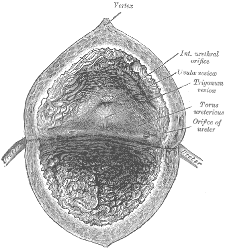
The bladder solves a problem that no other hollow organ faces. It has to expand from a few millilitres to half a litre without letting its internal pressure rise, hold a fluid that would poison the tissues around it if any of it leaked back, and then empty completely, on command, in a coordinated burst. Every distinctive feature of the organ follows from those three demands: a muscle arranged as a mesh rather than in layers, the least permeable epithelium in the body, and a reflex whose coordinating centre sits in the brainstem rather than in the spinal cord.
Anatomy: the structure
The empty bladder is a tetrahedral muscular sac lying entirely within the lesser pelvis, immediately behind the pubic symphysis; as it fills it becomes ovoid and rises into the abdomen, and in infants it is an abdominal organ from the start. It is described as having an apex pointing forward at the upper edge of the symphysis, a superior surface, two inferolateral surfaces, a posterior base or fundus, and a neck at the lowest point where the urethra begins. The median umbilical ligament, the fibrous remnant of the urachus, runs from the apex to the umbilicus. Peritoneum covers only the superior surface, and as the bladder fills it strips that peritoneum upward off the anterior abdominal wall, which is what makes suprapubic catheterisation of a distended bladder possible without entering the peritoneal cavity.
The interior is thrown into folds called rugae everywhere except over the trigone, a smooth triangular area on the base bounded by the two ureteric orifices and the internal urethral orifice. The trigone is smooth because its mucosa is firmly bound to the muscle beneath, and it is embryologically distinct, derived from absorbed mesonephric duct rather than from the endodermal urogenital sinus that forms the rest. A raised interureteric ridge joins the two ureteric orifices. The ureters do not enter perpendicularly; each passes obliquely through the bladder wall for 1.5 to 2 cm, so that rising intravesical pressure compresses the intramural segment against the muscle and closes it, a flap valve that prevents urine refluxing back toward the kidney. At the neck, smooth muscle forms the internal urethral sphincter, and further distally a striated external urethral sphincter in the deep perineal pouch provides voluntary control.
Relations differ between the sexes and this determines both symptoms and examination. In men the base is related to the seminal vesicles and vasa deferentia, with the prostate immediately below the neck and the rectum behind; in women the base rests directly on the anterior wall of the vagina, with the uterus lying on the superior surface. Arterial supply is from superior and inferior vesical branches of the internal iliac artery, with additional vaginal or middle rectal contributions. A rich vesical venous plexus drains into the internal iliac veins, and lymph passes to the external and internal iliac nodes. Innervation comes from three sources: parasympathetic pelvic splanchnic fibres from S2 to S4, which contract the detrusor; sympathetic fibres from T11 to L2 through the hypogastric nerves, which relax the detrusor and close the neck; and the somatic pudendal nerve from S2 to S4, which supplies the external sphincter. Sensory fibres run back along all three.
Physiology: how it works
Storage is an active achievement, not a passive one. As the bladder fills, pressure rises only a few centimetres of water across hundreds of millilitres, because the detrusor is viscoelastic and because noradrenaline acting on beta-3 receptors keeps it relaxed while sympathetic alpha-1 activity tightens the neck. At the same time the guarding reflex progressively increases pudendal firing to the external sphincter, so continence tightens as the bladder fills. First sensation of filling usually appears around 150 to 250 mL, a strong desire to void around 400 to 500 mL, and a healthy bladder holds that volume at a pressure below about 10 to 15 cmH2O. After voiding, residual volume is normally under 50 mL.
Voiding reverses all of this in a coordinated sequence, and the coordination happens in the brainstem. Stretch signals travel in the pelvic nerves to the sacral cord and ascend to the periaqueductal grey, which relays to the pontine micturition centre in Barrington nucleus. When voiding is permitted, that centre simultaneously drives sacral parasympathetic outflow, contracting the detrusor through M3 muscarinic receptors, and inhibits the pudendal motor neurons in Onuf nucleus, relaxing the external sphincter. Because detrusor contraction stimulates further afferent firing, the reflex is self-reinforcing and runs to completion once triggered. The frontal cortex holds a veto over the whole loop, which is why micturition is described as a spinobulbospinal reflex under voluntary control rather than a simple spinal reflex, and why a lesion between the pons and the sacral cord uncouples the muscle from its sphincter.
The lining does two jobs at once. As a barrier, the urothelium holds the steep gradients of urea, ammonia, potassium, and protons between urine and blood, and it is the least permeable epithelium in the body; the glycosaminoglycan layer on its surface adds a further defence against adherence and irritation. As a sensor, it is mechanically responsive: stretching releases ATP onto P2X3 receptors on suburothelial afferent nerve endings, along with acetylcholine and nitric oxide, so the epithelium itself participates in generating the sensation of fullness rather than merely lining the organ. The apical surface area is not fixed either. Discoidal and fusiform vesicles are inserted into the apical membrane as the bladder fills and retrieved as it empties, so the surface expands and contracts with the organ instead of stretching.
Biology: the living cells
The superficial cells of the urothelium, called umbrella cells, are the specialists. They are large, frequently binucleate and polyploid, and their apical membrane is an asymmetric unit membrane built from hexagonal plaques of the four uroplakins Ia, Ib, II, and IIIa, which together cover about 90 percent of the luminal surface and are the structural basis of the permeability barrier. Neighbouring umbrella cells are sealed by tight junctions rich in claudin-4 and claudin-8, and the whole layer turns over slowly, over a matter of months, yet regenerates rapidly after injury from the intermediate and basal cells beneath it. Basal cells express keratin 5 and p63 and act as the progenitor compartment. Beneath the epithelium, detrusor smooth muscle cells carry M3 muscarinic receptors for contraction and beta-3 adrenoceptors for relaxation, are electrically coupled by gap junctions so that activity spreads across the wall, and use Rho-kinase mediated calcium sensitisation to sustain tone economically. Between the two sit suburothelial myofibroblasts, sometimes called interstitial cells, which respond to urothelial ATP and are thought to act as intermediaries between the sensing surface and the afferent nerves. The pharmacology of every drug used for overactive bladder maps directly onto this list of receptors.
Histology: under the microscope
Four layers make up the wall, and the epithelium is the one that identifies it. The mucosa is lined by urothelium, also called transitional epithelium, which appears five to seven cells thick and rounded when the bladder is relaxed and only two or three cells thick and flattened when it is distended, so the same tissue looks different in two sections from the same organ. Its surface cells are the dome-shaped umbrella cells, with a scalloped luminal border, and there are no goblet cells and no keratin. Beneath it lies a lamina propria of dense connective tissue carrying elastic fibres, vessels, and nerves, often containing a discontinuous and incomplete muscularis mucosae, whose presence is a practical problem for pathologists because it can be mistaken for the true muscle layer. There is no distinct submucosa. The muscularis propria is the detrusor: three poorly demarcated and freely interlacing layers, described as inner longitudinal, middle circular, and outer longitudinal, which become clearly separate only at the bladder neck and around the ureteric orifices. Outside it is adventitia, replaced by serosa over the superior surface alone. The ureter above is distinguished by a much thinner wall and a star-shaped lumen, and the trigone by a smooth mucosa without rugae.
Clinical correlations
Urinary tract infection is the commonest bladder disease and its mechanism is anatomical and molecular together. The female urethra is short and close to the perineum, so ascending infection by uropathogenic Escherichia coli is easy; those bacteria then attach through type 1 fimbriae, whose FimH adhesin binds the mannosylated uroplakin Ia on the umbrella cell surface. Some invade the umbrella cells and form intracellular bacterial communities that survive antibiotic exposure and reseed the lumen later, which is one explanation for recurrent infection in the absence of reinfection from outside. Disruption of the surface glycosaminoglycan layer is implicated in interstitial cystitis, or bladder pain syndrome, in which pain and frequency persist without infection.
Obstruction and storage disorders show what happens when the mechanics fail. Benign prostatic enlargement obstructs the outlet, and the detrusor hypertrophies against it, producing the trabeculated wall and mucosal diverticula seen at cystoscopy, then incomplete emptying, rising residual volume, and eventually back-pressure on the ureters and kidneys. Overactive bladder is the opposite problem, involuntary detrusor contraction during filling, treated by blocking the M3 receptor or stimulating the beta-3 receptor. Neurological lesions map onto the reflex arc: a lesion above the pons removes voluntary inhibition and produces urgency with coordinated voiding; a spinal cord lesion between the pons and the sacral segments leaves the reflex intact but uncoordinated, so the detrusor contracts against a closed sphincter in detrusor-sphincter dyssynergia, generating high pressures that threaten the kidneys; a sacral or cauda equina lesion abolishes the reflex entirely, leaving a flaccid bladder with overflow incontinence.
More than 90 percent of bladder cancers are urothelial carcinomas, and the dominant risk factor is cigarette smoking, followed by occupational exposure to aromatic amines in the dye and rubber industries. Chronic infection with Schistosoma haematobium instead drives squamous metaplasia and squamous cell carcinoma, which is why the histological type differs by region. The classic presentation is painless visible haematuria, and any episode in an adult warrants investigation. Staging turns almost entirely on one question: whether the tumour has invaded the muscularis propria, since non-muscle-invasive disease can be managed with local resection and intravesical therapy while muscle-invasive disease requires radical treatment. That is why the distinction between a wisp of muscularis mucosae in the lamina propria and true detrusor muscle in a biopsy specimen carries so much weight. Because the whole urothelium is exposed to the same carcinogens, disease is often multifocal and recurrent, and surveillance cystoscopy is part of standard follow-up.
Common questions about the bladder
How much urine can the bladder hold before you need to go?
First awareness of filling usually appears at about 150 to 250 mL, a definite urge at around 400 to 500 mL, and most adults can hold 500 to 600 mL comfortably. What matters more than volume is pressure: a healthy bladder accommodates all of that while staying below roughly 10 to 15 cmH2O, and after voiding it should retain less than about 50 mL.
What is transitional epithelium and why does the bladder need it?
Transitional epithelium, or urothelium, is a stratified epithelium whose cells change shape as the organ expands, appearing several layers thick and rounded when relaxed and fewer layers thick and flattened when stretched. The bladder needs it because it combines that mechanical flexibility with an almost impermeable apical surface, so urine can be stored for hours without its solutes leaking back into the tissue.
What controls urination?
Stretch receptors in the bladder wall signal to the sacral spinal cord and then up to the pontine micturition centre, which coordinates the two halves of voiding: parasympathetic contraction of the detrusor and simultaneous relaxation of the external sphincter. The frontal cortex decides whether that centre is allowed to fire, which is how voiding stays voluntary in an adult.
Why are bladder infections more common in women?
The female urethra is much shorter and its opening lies closer to the perineum, so bacteria have a far shorter distance to travel to reach the bladder. Once there, uropathogenic Escherichia coli bind to uroplakin on the surface cells using their type 1 fimbriae, and some persist inside those cells, which contributes to infections that recur rather than simply returning from outside.
Selected references
- Standring S, ed. Gray's Anatomy: The Anatomical Basis of Clinical Practice. 42nd ed. Elsevier; 2021.
- Ross MH, Pawlina W. Histology: A Text and Atlas. 8th ed. Wolters Kluwer; 2020.
- Hall JE, Hall ME. Guyton and Hall Textbook of Medical Physiology. 14th ed. Elsevier; 2021.
- Fowler CJ, Griffiths D, de Groat WC. The neural control of micturition. Nature Reviews Neuroscience. 2008;9(6):453-466.
- Khandelwal P, Abraham SN, Apodaca G. Cell biology and physiology of the uroepithelium. American Journal of Physiology - Renal Physiology. 2009;297(6):F1477-F1501.
- Kumar V, Abbas AK, Aster JC. Robbins and Cotran Pathologic Basis of Disease. 10th ed. Elsevier; 2021.
Medical disclaimer. This article is written for education and general understanding. It is not medical advice and cannot replace assessment by a qualified healthcare professional. If you have symptoms or health concerns, speak with a clinician. Read the full disclaimer.