15 studies
Choose an organ. Follow the system.
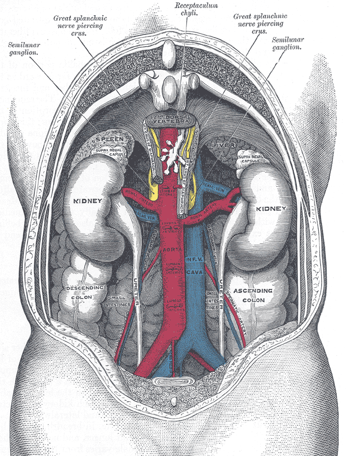
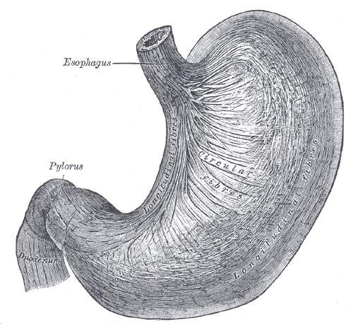
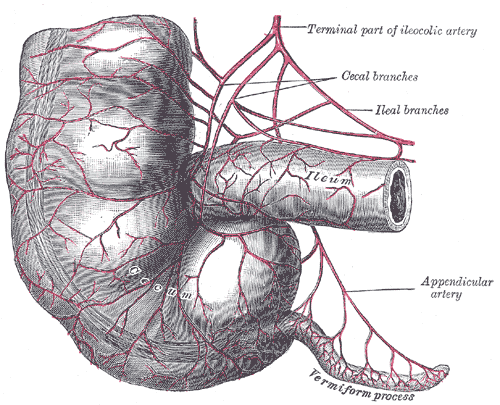
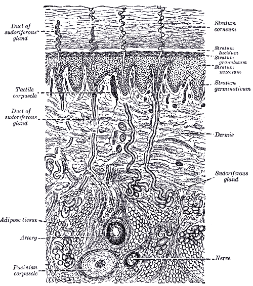
Fifteen organs, each one traced through the same four lenses in the same order: where it sits and what it is attached to, how it works, the cells that do the work, and what the tissue looks like under a microscope. Every guide ends with the clinical picture — what goes wrong, and why the anatomy predicts it.
Start here
New to this section? These three are the ones to read first, in this order.
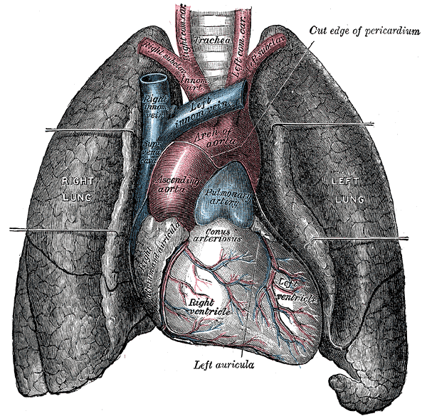
- 01HeartA complete guide to the human heart: chambers, valves and coronary vessels, the cardiac cycle, excitation-contraction coupling, cardiomyocyte biology, and the histology of all three cardiac layers.
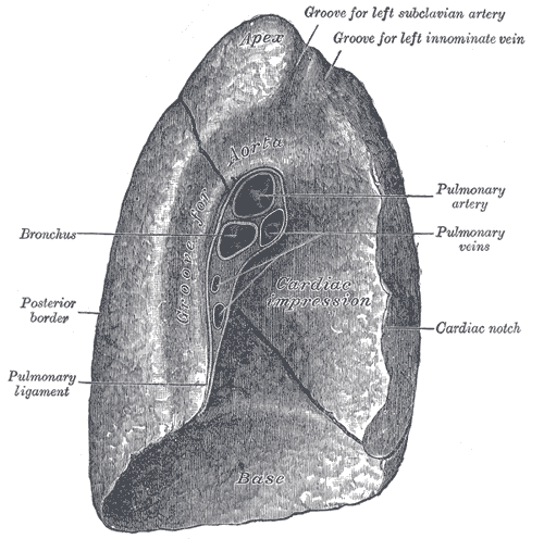
- 02LungsHow the lungs are built and how they work: the bronchial tree, pleura and respiratory mechanics, gas exchange and oxygen transport, alveolar cell biology, and the histology of the respiratory membrane.
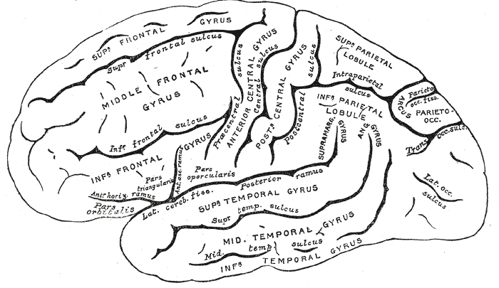
- 03BrainA structured guide to the human brain: lobes and deep structures, the meninges and cerebrospinal fluid, synaptic transmission, neuronal and glial biology, and the histology of grey and white matter.
Every study in this section
Every major organ traced from its position in the body down to the cells that make it work.