The nervous system is the body's fast control network, and speed is its entire justification. Hormones can carry a message anywhere in the body, but they take seconds to minutes and cannot be addressed to one muscle. A nerve impulse reaches a specific target in milliseconds. The system that does this is usually split three ways — central and peripheral by location, somatic and autonomic by what they control, sympathetic and parasympathetic within the autonomic branch — and the divisions overlap, which is why they confuse people. They are three different questions asked about the same wiring.
The parts, and how they fit together
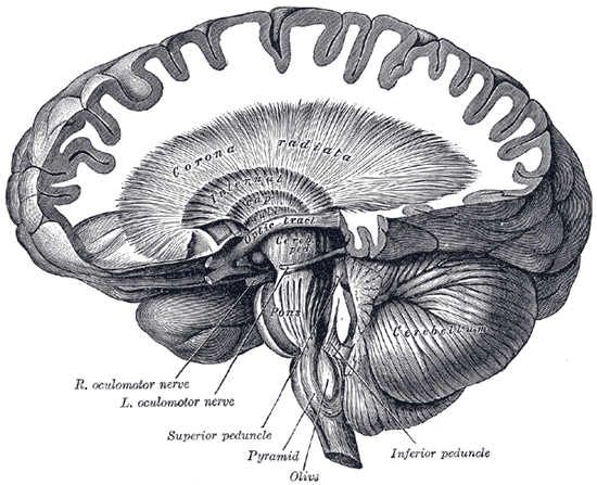
The first division is anatomical. The central nervous system is the brain and spinal cord, protected by bone, wrapped in three meninges — dura, arachnoid and pia — and cushioned by cerebrospinal fluid in the subarachnoid space and ventricles. The peripheral nervous system is everything else: 12 pairs of cranial nerves emerging from the brain and brainstem, and 31 pairs of spinal nerves emerging from the cord, each formed by a dorsal root carrying sensory fibres in and a ventral root carrying motor fibres out. Cell bodies gather into nuclei inside the central nervous system and into ganglia outside it, and the same distinction applies to the cells that insulate axons: oligodendrocytes centrally, Schwann cells peripherally.
The second division is functional. The somatic nervous system handles conscious sensation and voluntary movement of skeletal muscle, using a single motor neuron running all the way from the spinal cord to the muscle, releasing acetylcholine, and always excitatory. The autonomic nervous system handles everything you do not think about — heart rate, blood vessel diameter, airway calibre, gut motility, glands, pupils — using a two-neuron chain with a synapse in a ganglion on the way, and it can be excitatory or inhibitory depending on the receptor.
The autonomic system splits again. The sympathetic division has short preganglionic fibres leaving the cord between the first thoracic and second lumbar segments, synapsing in the nearby sympathetic chain, and long postganglionic fibres releasing noradrenaline — an arrangement that lets one signal spread widely and produce a whole-body response. The parasympathetic division has long preganglionic fibres leaving through cranial nerves III, VII, IX and X and through sacral segments two to four, synapsing in ganglia close to or within the target organ, and short postganglionic fibres releasing acetylcholine, which allows precise organ-by-organ control. The vagus nerve alone carries about three-quarters of all parasympathetic output. A third division, the enteric nervous system in the gut wall, holds several hundred million neurons and runs largely independently.
How the system works
The unit of signalling is the action potential, and it is all-or-nothing. A neuron at rest holds its inside at around minus 70 millivolts, maintained by the sodium-potassium pump and by the membrane being far more permeable to potassium than sodium. Depolarisation to threshold opens voltage-gated sodium channels, sodium floods in and drives the membrane to about plus 30 millivolts, those channels then inactivate while potassium channels open and restore the resting state. The whole event takes about a millisecond, and because the size never varies, information is carried in firing frequency rather than amplitude. Myelin makes it fast: current flows passively down insulated internodes and the impulse regenerates only at the nodes of Ranvier, reaching up to 120 metres per second against about one in unmyelinated fibres.
Decisions are made by summation. A single neuron may receive thousands of synaptic inputs, each producing a small excitatory or inhibitory change in membrane potential; those changes add together in space and time, and the neuron fires only if the total reaches threshold at the axon hillock. Signals cross synapses chemically: the arriving action potential opens calcium channels, vesicles of neurotransmitter fuse with the membrane, and receptors on the next cell convert the chemical signal back to an electrical one. The transmitter identifies the pathway — acetylcholine at every neuromuscular junction and all autonomic ganglia, noradrenaline at most sympathetic targets, glutamate as the main central excitatory transmitter, GABA and glycine as the main inhibitory ones, and dopamine, serotonin and others as modulators.
The simplest complete circuit is the reflex arc, and it explains why some responses are faster than thought. A stretch receptor in a muscle spindle sends a sensory neuron into the spinal cord, which synapses directly onto the motor neuron for the same muscle; the muscle contracts before the signal has reached the brain at all. Adding one interneuron produces the polysynaptic withdrawal reflex that pulls a hand from something hot. At the other extreme, the sympathetic and parasympathetic divisions run continuous background tone rather than switching on and off, so heart rate is set by the balance between them. Sympathetic activation raises heart rate and contractility, dilates pupils and bronchi, redirects blood from gut to muscle, and mobilises glucose; parasympathetic activation slows the heart, constricts pupils and bronchi, and promotes digestion and bladder emptying. Most organs receive both, and the resting balance is parasympathetic — which is why cutting the vagus makes the heart speed up.
The cells that do the work
Nervous tissue is two populations with very different capabilities. Neurons carry the signals: a cell body with a pale nucleus, prominent nucleolus and coarse Nissl substance of rough endoplasmic reticulum; dendrites collecting input across an enormous surface; and one axon, which may be a metre long, carrying output. They are classified by process count — multipolar for most central neurons, bipolar in retina and olfactory epithelium, pseudounipolar for the sensory neurons whose cell bodies sit in dorsal root ganglia. With very few exceptions they leave the cell cycle permanently and cannot be replaced. Glial cells outnumber them and do everything else: astrocytes buffer potassium and neurotransmitter, supply neurons with lactate, and induce the tight junctions that create the blood-brain barrier; oligodendrocytes myelinate up to fifty central axons each; microglia act as resident immune cells; ependymal cells line the ventricles and circulate cerebrospinal fluid; Schwann cells myelinate a single peripheral internode each. That last difference has a large clinical consequence: a Schwann cell survives injury and leaves an intact basal lamina tube that guides a regrowing axon back at roughly a millimetre a day, whereas central axons meet inhibitory myelin debris and a glial scar and do not regrow.
Under the microscope
Grey matter and white matter look almost like different tissues. In grey matter, neuronal cell bodies are the obvious feature — large, often pyramidal, with a strikingly pale round nucleus and a single dark nucleolus like an owl's eye, and coarse basophilic Nissl clumps that fill the soma and dendrites but stop abruptly at the axon hillock, which is how you tell which process is the axon. Scattered densely between them are far more numerous small dark nuclei with almost no visible cytoplasm: the glia, which routine staining cannot separate into types. The pale pink felted background is the neuropil, a mat of dendrites, axons and glial processes. White matter looks nearly empty by comparison, rows of glial nuclei between pale tracts, because the myelin lipid dissolves during processing and leaves axons as ghostly circles or lines. A peripheral nerve in cross-section shows bundles of axons each surrounded by endoneurium, grouped into fascicles by perineurium, and bound together by epineurium — the same three-sheath arrangement as muscle, and the reason a nerve can be repaired surgically by suturing sheaths rather than axons.
When the system fails
Where a lesion sits determines what is lost, and the somatic system makes this especially predictable. Upper motor neuron lesions, in the brain or cord above the anterior horn, produce weakness with increased tone, brisk reflexes and an upgoing plantar response, because the descending inhibition on spinal reflexes is removed. Lower motor neuron lesions, from anterior horn cell to muscle, produce weakness with reduced tone, absent reflexes, wasting and fasciculation. Spinal cord injury cuts everything crossing that level, so the level itself predicts the deficit — and because the phrenic nerve arises from cervical roots three to five, injury above that stops spontaneous breathing.
Demyelination attacks the insulation rather than the wire. Multiple sclerosis destroys central myelin in plaques scattered through brain, optic nerves and cord; because oligodendrocytes remyelinate poorly, deficits accumulate, and the lesions are separated in both time and place. Guillain-Barré syndrome is the peripheral counterpart, usually following an infection, producing ascending weakness that starts in the legs — but because Schwann cells remyelinate well, most patients recover substantially. Myasthenia gravis attacks neither, targeting the acetylcholine receptor at the neuromuscular junction and producing weakness that worsens with repeated use.
Autonomic pharmacology follows directly from the anatomy, which is why it is worth learning once properly. Beta-blockers slow the heart by blocking sympathetic beta-1 receptors; beta-2 agonists dilate bronchi in asthma; alpha-1 blockers relax vascular and prostatic smooth muscle. Anticholinergics dry secretions, dilate pupils and speed the heart by removing parasympathetic tone, which also explains their side effect profile. Autonomic failure itself presents as postural hypotension, impotence, bladder dysfunction and gastroparesis, most commonly in long-standing diabetes and in Parkinson's disease. And because neurons cannot be replaced, the neurodegenerative diseases produce deficits in the pattern dictated by which population dies: substantia nigra in Parkinson's, cortex and hippocampus in Alzheimer's, motor neurons in amyotrophic lateral sclerosis.
Common questions about the nervous system
What is the difference between the central and peripheral nervous systems?
The central nervous system is the brain and spinal cord, encased in bone and wrapped in meninges. The peripheral nervous system is everything outside it: 12 pairs of cranial nerves, 31 pairs of spinal nerves, and the ganglia along them. The distinction matters clinically because peripheral axons regenerate at about a millimetre a day thanks to Schwann cells, while central axons essentially do not.
What is the difference between the sympathetic and parasympathetic nervous systems?
Both are autonomic and mostly act on the same organs in opposite directions. Sympathetic activity prepares for exertion: faster heart, dilated pupils and bronchi, blood diverted from gut to muscle, glucose mobilised. Parasympathetic activity supports rest and digestion: slower heart, constricted pupils, increased gut motility and secretion. Sympathetic fibres release noradrenaline at the target and leave the cord between T1 and L2; parasympathetic fibres release acetylcholine and leave via cranial nerves III, VII, IX and X and sacral segments S2 to S4.
What is a reflex arc?
The shortest complete circuit in the nervous system: a receptor, a sensory neuron, an integrating centre in the spinal cord, a motor neuron, and an effector. In a monosynaptic stretch reflex the sensory neuron synapses directly onto the motor neuron, so the muscle contracts before the brain is even informed. Adding one interneuron gives the withdrawal reflex that pulls your hand off something hot. Testing reflexes is useful precisely because it isolates one spinal level.
How fast do nerve signals travel?
It depends almost entirely on myelination and diameter. Large myelinated fibres conduct at up to about 120 metres per second, because the impulse jumps between nodes of Ranvier rather than travelling continuously. Small unmyelinated fibres, such as those carrying slow burning pain and most autonomic output, conduct at around one metre per second. That difference is why you feel the sharp part of an injury well before the dull ache.
Selected references
- Kandel ER, Koester JD, Mack SH, Siegelbaum SA. Principles of Neural Science. 6th ed. McGraw Hill; 2021.
- Standring S, ed. Gray's Anatomy: The Anatomical Basis of Clinical Practice. 42nd ed. Elsevier; 2021.
- Hall JE, Hall ME. Guyton and Hall Textbook of Medical Physiology. 14th ed. Elsevier; 2021.
- Azevedo FA, Carvalho LR, Grinberg LT, et al. Equal numbers of neuronal and nonneuronal cells make the human brain an isometrically scaled-up primate brain. Journal of Comparative Neurology. 2009;513(5):532-541.
- Abbott NJ, Patabendige AA, Dolman DE, Yurdakul SR, Begley DJ. Structure and function of the blood-brain barrier. Neurobiology of Disease. 2010;37(1):13-25.
- Kumar V, Abbas AK, Aster JC. Robbins and Cotran Pathologic Basis of Disease. 10th ed. Elsevier; 2021.
Medical disclaimer. This article is written for education and general understanding. It is not medical advice and cannot replace assessment by a qualified healthcare professional. If you have symptoms or health concerns, speak with a clinician. Read the full disclaimer.