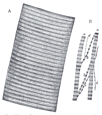
There are only three muscle tissues in the human body, and every movement you have ever made — lifting a cup, pushing food along the gut, ejecting blood from the heart — is one of them shortening. They use the same molecular trick: actin and myosin filaments dragging past each other, powered by ATP and switched on by calcium. What differs is who gives the order, how the filaments are arranged, and how fast the tissue can go from rest to full force and back. Those three differences explain everything else about them, including why only one of the three can be tired out and only one of the three cannot be repaired.
Where it is, and what it looks like
Skeletal muscle is attached to bone by tendon, is under voluntary control through somatic motor nerves, and makes up around 40 per cent of adult body mass. Its unit is the muscle fibre, a single cell formed by the fusion of many myoblasts during development, which is why it carries dozens or hundreds of nuclei pushed to the periphery just under the cell membrane. Fibres are bundled into fascicles by connective tissue sheaths — endomysium around each fibre, perimysium around each fascicle, epimysium around the whole muscle — which merge at the ends to form the tendon, so force generated anywhere in the belly is transmitted to bone.
Smooth muscle lines the walls of hollow organs: the gut, the airways, the bladder, the uterus, and every blood vessel larger than a capillary. It is involuntary, driven by autonomic nerves, hormones, local metabolites and stretch, and it usually forms sheets rather than bundles — typically an inner circular and an outer longitudinal layer, which is the arrangement that makes peristalsis possible. Each cell is a single spindle-shaped unit, 20 to 200 micrometres long, tapering at both ends, with one central nucleus that visibly corkscrews when the cell contracts.
Cardiac muscle exists only in the heart and in the roots of the great veins where they enter it. It is involuntary but, unlike smooth muscle, it generates its own rhythm from pacemaker cells rather than waiting for a signal. Its cells are short, branched and striated, usually with one central nucleus, and they are joined end to end at intercalated discs — specialised junctions that combine desmosomes and fascia adherens for mechanical continuity with gap junctions for electrical continuity. That combination lets the whole myocardium contract as a single coordinated unit, a functional syncytium, from one electrical trigger.
What it does
The shared mechanism is the sliding filament model. Thin filaments of actin and thick filaments of myosin interdigitate; when calcium becomes available, myosin heads bind actin, pivot, and pull the thin filaments toward the centre, then detach using energy from ATP and repeat. The filaments themselves never shorten — they slide past one another, and the tissue shortens because the overlap increases. Relaxation is not passive: calcium has to be actively pumped back out of the cytoplasm against its gradient, which is why rigor mortis appears when ATP production stops and the cross-bridges can neither detach nor be released.
What differs is the switch. In skeletal and cardiac muscle, calcium binds troponin C on the thin filament, which shifts tropomyosin off the myosin binding sites — the control is on the actin. Skeletal muscle releases that calcium from its own sarcoplasmic reticulum in response to membrane depolarisation alone, so it can contract in a few milliseconds and is fully independent of extracellular calcium. Cardiac muscle needs a small influx of calcium through L-type channels to trigger a much larger release from the sarcoplasmic reticulum, which is calcium-induced calcium release, and it makes cardiac force adjustable by anything that changes calcium entry. Smooth muscle controls the myosin instead: calcium binds calmodulin, which activates myosin light chain kinase, which phosphorylates myosin and allows it to engage actin at all.
Those differences set the tempo. Skeletal muscle is fast, precise and gradable by recruiting more motor units and firing them faster, and because a single action potential is much shorter than the resulting twitch it can be summed into a sustained tetanic contraction — and can fatigue. Cardiac muscle cannot be tetanised: the plateau phase of its action potential holds the cell refractory for roughly 200 milliseconds, almost as long as the contraction itself, which guarantees the heart relaxes and refills between beats. Smooth muscle is slow, cheap and tireless: it develops force over seconds rather than milliseconds, and its latch mechanism lets it maintain tension for hours on a fraction of the ATP skeletal muscle would need, which is what holds a blood vessel at a set diameter all day.
The cells that build it
The contractile machinery is built into repeating units called sarcomeres in skeletal and cardiac muscle, and the striations you see are simply those units in register across the whole cell. Each sarcomere runs from one Z line to the next and contains a dark A band of thick myosin filaments and a light I band of thin actin filaments, held in alignment by titin, the largest protein in the body, which acts as a molecular spring and sets passive tension. Around this sit the regulatory proteins troponin and tropomyosin, and the structural proteins that tie the machinery to the cell membrane, of which dystrophin is the one that matters clinically. Smooth muscle has actin and myosin too, but arranged obliquely and anchored to dense bodies scattered through the cytoplasm rather than to Z lines, which is why it has no striations and why it can shorten far more of its resting length than skeletal muscle can. The cells also differ in what they can replace. Skeletal muscle keeps a reserve of satellite cells beneath the basal lamina that can proliferate and fuse into damaged fibres, so it repairs well. Smooth muscle cells retain the ability to divide outright, which is how the uterus grows in pregnancy and how arteries thicken in hypertension. Cardiomyocytes renew at well under one per cent per year, which is far too slow to matter after an infarct, so dead myocardium is replaced by scar.
How to recognise it on a slide
Three questions separate the three tissues on any slide. First: are there striations? If the cytoplasm shows regular cross-bands, it is skeletal or cardiac; if it is smooth and evenly pink, it is smooth muscle. Second: how many nuclei, and where are they? Skeletal fibres are multinucleate with the nuclei flattened against the edge of the cell, immediately under the membrane. Cardiac and smooth muscle cells each have one central nucleus. Third: do the fibres branch? Skeletal fibres are long, straight, parallel cylinders that never branch and are often enormous — up to several centimetres long. Cardiac fibres branch and rejoin in a network, and are crossed by dark transverse lines, the intercalated discs, which are the single most reliable feature of cardiac muscle and are found nowhere else in the body. Smooth muscle in longitudinal section looks like overlapping spindles with cigar-shaped nuclei, and in transverse section like a field of circles of very different diameters — because a spindle cut near its tapered end is small and has no nucleus in the plane of section, while one cut through its middle is wide and does.
When it goes wrong
The muscular dystrophies show what happens when the link between the contractile machinery and the cell membrane fails. In Duchenne muscular dystrophy, dystrophin is absent, so each contraction tears the sarcolemma slightly; fibres degenerate, satellite cells exhaust themselves trying to regenerate them, and muscle is progressively replaced by fat and fibrous tissue. It presents in early childhood with difficulty rising from the floor and a characteristic waddling gait, and creatine kinase is grossly elevated because the damaged fibres leak it into the blood. Becker muscular dystrophy is the same protein made badly rather than not at all, and runs a milder course.
Acute muscle breakdown produces rhabdomyolysis, in which myoglobin released from crushed or over-exerted fibres reaches the kidneys and precipitates in the tubules, causing acute kidney injury. Causes range from crush injury and prolonged immobility to extreme exercise, seizures, certain drugs and statins. Myasthenia gravis attacks the junction rather than the muscle: antibodies against the acetylcholine receptor at the neuromuscular junction reduce the signal reaching an otherwise healthy fibre, producing weakness that characteristically worsens through the day and with repeated use.
Smooth and cardiac muscle fail in ways that reflect what they normally do. Bronchial smooth muscle constriction and hypertrophy is central to asthma, which is why bronchodilators target beta-2 receptors on those cells. Vascular smooth muscle tone sets peripheral resistance, so most antihypertensive drugs work either on it or on the signals that reach it, and its proliferation thickens arterial walls in chronic hypertension. In the heart, loss of cardiomyocytes after a myocardial infarction is permanent, because the scar that replaces them can neither contract nor conduct — which is why heart failure after infarction is progressive and why arrhythmias so often arise at the scar border.
Common questions about muscle tissue
What is the difference between skeletal, smooth, and cardiac muscle?
Skeletal muscle is voluntary, striated, attached to bone, and made of long unbranched multinucleate fibres with peripheral nuclei. Cardiac muscle is involuntary, striated, found only in the heart, and made of short branching cells with one central nucleus joined by intercalated discs. Smooth muscle is involuntary, has no striations, lines hollow organs and vessels, and is made of single spindle-shaped cells with one central nucleus.
How can you tell cardiac muscle from skeletal muscle on a slide?
Both are striated, so use branching and nuclei instead. Cardiac fibres branch and rejoin, carry a single central nucleus, and are crossed by dark intercalated discs. Skeletal fibres run in straight parallel lines without branching and have many nuclei squeezed against the outside edge of the cell. Intercalated discs are the decider, since nothing else in the body has them.
Why does smooth muscle have no striations?
Because its actin and myosin are not arranged in sarcomeres. Instead of being lined up in repeating units between Z lines, the filaments run obliquely across the cell and anchor to dense bodies scattered through the cytoplasm and along the membrane. There is no regular repeating pattern to produce a banded appearance, and the oblique arrangement is also why smooth muscle can shorten far more of its resting length than striated muscle can.
Which types of muscle can regenerate?
Skeletal muscle repairs well, using satellite cells that sit under the basal lamina and fuse into damaged fibres. Smooth muscle cells can divide outright, which is how the uterus enlarges in pregnancy. Cardiac muscle effectively cannot: adult cardiomyocytes renew at well under one per cent a year, so tissue lost in a heart attack is replaced by collagen scar and the function is not recovered.
Selected references
- Ross MH, Pawlina W. Histology: A Text and Atlas. 8th ed. Wolters Kluwer; 2020.
- Hall JE, Hall ME. Guyton and Hall Textbook of Medical Physiology. 14th ed. Elsevier; 2021.
- Mescher AL. Junqueira's Basic Histology: Text and Atlas. 16th ed. McGraw Hill; 2021.
- Bers DM. Cardiac excitation-contraction coupling. Nature. 2002;415(6868):198-205.
- Webster C, Silberstein L, Hays AP, Blau HM. Fast muscle fibers are preferentially affected in Duchenne muscular dystrophy. Cell. 1988;52(4):503-513.
- Kumar V, Abbas AK, Aster JC. Robbins and Cotran Pathologic Basis of Disease. 10th ed. Elsevier; 2021.
Medical disclaimer. This article is written for education and general understanding. It is not medical advice and cannot replace assessment by a qualified healthcare professional. If you have symptoms or health concerns, speak with a clinician. Read the full disclaimer.