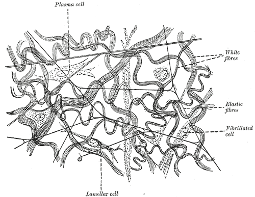
Every other tissue in the body is defined by its cells. Connective tissue is defined by what its cells have built and then moved out of the way of. Its properties — the tension a tendon holds, the spring in an artery wall, the hardness of bone, the fluidity of blood — come almost entirely from the extracellular matrix, which is why a single tissue class can include something as rigid as the femur and something as liquid as plasma. Once you read connective tissue as a recipe of three ingredients in different proportions, the whole classification stops being a list to memorise.
Where it is, and what it looks like
The three ingredients are cells, fibres and ground substance, and the last two together make the extracellular matrix. Connective tissue proper is divided by how densely those fibres are packed. Loose, or areolar, connective tissue has fibres running in every direction with plenty of space between them; it is the packing material of the body, filling the gaps around organs, underlying every epithelium as the lamina propria, and carrying the small vessels and nerves that supply the tissue above. Adipose tissue is loose connective tissue in which fat cells have crowded out almost everything else. Reticular tissue, a mesh of fine type III collagen, forms the internal scaffold of the spleen, lymph nodes, liver and bone marrow.
Dense connective tissue has the same ingredients with far more collagen and much less ground substance, and it is subdivided by whether that collagen is organised. Dense regular connective tissue runs its fibres in parallel bundles along one axis, producing enormous tensile strength in that direction and very little in any other: this is tendon, which ties muscle to bone, and ligament, which ties bone to bone. Dense irregular connective tissue weaves its collagen in all directions, giving strength against pull from any angle, and forms the dermis of the skin, the capsules around organs, and the tough sheaths around nerves and bones.
Three specialised tissues sit inside the same family because they share the same basic recipe with an unusual matrix. In cartilage the ground substance is a firm gel, so the tissue is stiff but bendable and, uniquely, has no blood supply of its own. In bone the matrix is impregnated with calcium hydroxyapatite crystals, making it rigid enough to bear weight while remaining alive and constantly remodelled. In blood the matrix is liquid plasma and the fibres exist only in potential, appearing as fibrin when clotting is triggered. Lymph is the same idea again, drained from the tissue spaces rather than pumped.
What it does
The first job is mechanical, and it is done by the fibres. Collagen, the most abundant protein in the human body at roughly a third of total protein mass, resists tension: a collagen fibre has a tensile strength comparable to mild steel for its weight and stretches almost not at all, which is what lets a tendon transmit the full force of a muscle to a bone without lengthening. Elastic fibres do the opposite, stretching to around one and a half times their length and recoiling completely, which is what allows the aorta to expand with each heartbeat and return the stored energy to the blood during diastole. Reticular fibres form fine supporting nets rather than load-bearing cables.
The second job belongs to the ground substance, and it is transport. Ground substance is a viscous gel of glycosaminoglycans, proteoglycans and adhesive glycoproteins, and it holds a great deal of water. Every nutrient reaching an epithelial cell and every waste product leaving it crosses this gel by diffusion, so the connective tissue beneath an epithelium is not merely padding but the route through which the tissue above stays alive. Its water-binding capacity also makes it a shock absorber and explains oedema, which is simply too much fluid held in that same compartment.
The third job is defence and repair, and it is the reason loose connective tissue is where inflammation happens. It is already populated with macrophages, mast cells and wandering white cells, and its vessels are the ones that dilate and leak when tissue is injured or infected. When healing begins, fibroblasts migrate in, proliferate, and lay down new collagen, first as disorganised granulation tissue and then as progressively remodelled scar. Adipose tissue adds a fourth job that was underestimated for a century: it is the body's main energy store and also an endocrine organ, releasing leptin, adiponectin and inflammatory mediators that act on the brain, liver and muscle.
The cells that build it
The resident cell is the fibroblast, and almost everything a connective tissue is comes from what its fibroblasts have secreted. Fibroblasts synthesise procollagen, elastin, and the proteoglycans of the ground substance, then remain in the tissue as quiescent fibrocytes until injury reactivates them. Around them sit a standing population of defensive cells: macrophages, which phagocytose debris and present antigen; mast cells, packed with histamine and heparin granules and triggered by IgE; plasma cells, which are antibody factories derived from B lymphocytes; and adipocytes, each of which holds a single fat droplet so large that it flattens the nucleus against the cell membrane. The matrix they build is more specific than it looks. Collagen exists as at least 28 types, of which four cover most of the body: type I in bone, tendon, ligament, dermis and organ capsules; type II in hyaline and elastic cartilage; type III as the reticular fibres of lymphoid and haematopoietic organs; and type IV as the flat sheet of the basement membrane, which is the one type that does not form fibrils. Assembling type I collagen requires hydroxylation of proline and lysine residues, a reaction that cannot proceed without vitamin C — which is why scurvy is, at the cellular level, a connective tissue disease.
How to recognise it on a slide
Connective tissue is the mirror image of epithelium under the microscope: sparse cells with a great deal of material between them, and no free surface. In a haematoxylin and eosin section the collagen stains pink and the nuclei of the widely spaced fibroblasts appear as thin dark spindles lying along the fibres. Loose areolar tissue looks open and untidy, with strands of pink collagen running in every direction, obvious empty-looking spaces, and small vessels crossing the field. Dense regular tissue is unmistakable once seen: long parallel waves of pink collagen with rows of flattened fibroblast nuclei squeezed between the bundles like text between ruled lines. Dense irregular tissue has the same heavy pink collagen with no dominant direction. Adipose tissue looks like chicken wire, because the fat is dissolved out during processing and leaves large empty polygons with a thin rim of cytoplasm and a nucleus pushed to one edge. Hyaline cartilage shows chondrocytes sitting in lacunae within a smooth glassy blue-pink matrix; bone shows concentric lamellae around Haversian canals; and blood, of course, shows cells with no fibres at all until it clots.
When it goes wrong
Inherited faults in matrix proteins produce some of the clearest demonstrations in medicine that a tissue is only as good as what its cells secreted. In Ehlers-Danlos syndrome, defects in collagen synthesis or processing give hypermobile joints, skin that stretches and scars poorly, and, in the vascular type, arteries and hollow organs that can rupture. In Marfan syndrome the defective protein is fibrillin-1, the scaffold on which elastic fibres are built, producing tall stature, long limbs, lens dislocation and progressive dilatation of the aortic root. In osteogenesis imperfecta, type I collagen is faulty and bones fracture under ordinary loads.
Acquired connective tissue disease is more common still. Scurvy, from vitamin C deficiency, prevents collagen from being cross-linked properly, so old collagen degrades faster than new collagen can replace it — hence bleeding gums, poor wound healing and reopening of long-healed scars. In systemic sclerosis, fibroblasts are driven to overproduce collagen, thickening skin and eventually the lungs, gut and kidneys. Keloid scars are the same error confined to a wound. At the other extreme, fibrosis is the general term for connective tissue replacing functioning tissue after injury, and it is the final common pathway of chronic damage in the liver, lung, kidney and heart.
Because loose connective tissue is where inflammation and fluid collect, it also determines how infection and swelling spread. Cellulitis is infection of the loose connective tissue beneath the skin, and it spreads along tissue planes rather than respecting anatomical boundaries. Oedema is excess fluid in the ground substance, produced whenever capillary pressure rises, plasma protein falls, or lymphatic drainage fails. And adipose tissue, now understood as an endocrine organ, links obesity to insulin resistance and chronic low-grade inflammation through the mediators it secretes rather than through its bulk alone.
Common questions about connective tissue
What are the main types of connective tissue?
Connective tissue proper divides into loose forms — areolar, adipose and reticular — and dense forms, which are either regular, as in tendons and ligaments, or irregular, as in the dermis and organ capsules. Three specialised connective tissues complete the family: cartilage, with a firm gel matrix; bone, with a mineralised matrix; and blood, with a liquid one.
What is the difference between connective tissue and epithelium?
Epithelium is defined by its cells, which are packed tightly together on a basement membrane with almost no material between them, and it contains no blood vessels. Connective tissue is defined by its extracellular matrix: the cells are sparse and widely separated, most of the tissue is fibres and ground substance, and it is richly supplied with vessels. On a slide, crowded cells with a free surface mean epithelium; scattered cells in a pink fibrous background mean connective tissue.
What does a fibroblast do?
The fibroblast is the resident cell of connective tissue and it builds the matrix. It synthesises and secretes collagen, elastin and the proteoglycans of the ground substance, then settles into a quiet state as a fibrocyte. After injury it reactivates, migrates into the wound, multiplies and lays down new collagen, which is the basis of granulation tissue and of every scar.
Why is cartilage classed as connective tissue if it has no blood supply?
Because it follows the same recipe: scattered cells, chondrocytes, surrounded by a matrix they secreted themselves. What differs is the matrix, a firm gel of type II collagen and proteoglycan that is stiff enough to resist compression but too dense for vessels to grow through. Chondrocytes are fed by diffusion from the perichondrium instead, which is exactly why cartilage heals so slowly.
Selected references
- Ross MH, Pawlina W. Histology: A Text and Atlas. 8th ed. Wolters Kluwer; 2020.
- Mescher AL. Junqueira's Basic Histology: Text and Atlas. 16th ed. McGraw Hill; 2021.
- Standring S, ed. Gray's Anatomy: The Anatomical Basis of Clinical Practice. 42nd ed. Elsevier; 2021.
- Kumar V, Abbas AK, Aster JC. Robbins and Cotran Pathologic Basis of Disease. 10th ed. Elsevier; 2021.
- Ricard-Blum S. The collagen family. Cold Spring Harbor Perspectives in Biology. 2011;3(1):a004978.
- Kershaw EE, Flier JS. Adipose tissue as an endocrine organ. Journal of Clinical Endocrinology and Metabolism. 2004;89(6):2548-2556.
Medical disclaimer. This article is written for education and general understanding. It is not medical advice and cannot replace assessment by a qualified healthcare professional. If you have symptoms or health concerns, speak with a clinician. Read the full disclaimer.