The pancreas is two organs sharing one address. About 98 percent of its mass is exocrine tissue producing the most powerful digestive enzyme mixture in the body, and the remaining 2 percent, scattered through it as roughly a million islets of Langerhans, controls the fuel economy of every cell. Both functions are essential, both fail in recognisable ways, and both are best understood by starting from the tissue that performs them.
Anatomy: the structure
The pancreas lies retroperitoneally across the posterior abdominal wall at roughly the L1 to L2 level, behind the stomach. It is described in four parts: the head nestled in the C-shaped curve of the duodenum, with the uncinate process hooking behind the superior mesenteric vessels; the neck; the body crossing the midline; and the tail, the only part that is intraperitoneal, reaching into the splenorenal ligament toward the splenic hilum. This position explains why pancreatic pain often radiates to the back.
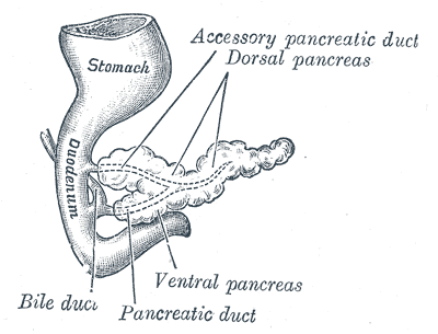
The main pancreatic duct, the duct of Wirsung, runs the length of the organ collecting side branches and joins the common bile duct at the ampulla of Vater, where the sphincter of Oddi controls release into the second part of the duodenum. An accessory duct of Santorini often persists and drains separately into the minor papilla. This shared final pathway with the biliary tree is the anatomical reason gallstones can cause pancreatitis.
Blood supply reflects the organ's foregut and midgut boundary. The head is supplied by superior and inferior pancreaticoduodenal arteries, arising from the gastroduodenal and superior mesenteric arteries respectively, forming arcades shared with the duodenum, which is why the two cannot easily be separated surgically. The body and tail are supplied by branches of the splenic artery. Venous drainage is into the portal system, so insulin released from islets reaches the liver at high concentration before entering the general circulation.
Physiology: how it works
The exocrine pancreas secretes about 1.5 litres of alkaline juice daily with two distinct components. Duct cells secrete bicarbonate under the influence of secretin, released when acid chyme enters the duodenum, raising duodenal pH toward the range where pancreatic enzymes work. Acinar cells secrete the enzymes themselves under the influence of cholecystokinin, released in response to fat and protein, and vagal stimulation. Together these cover every macronutrient: amylase for starch, lipase with colipase for triglycerides, and trypsinogen, chymotrypsinogen, procarboxypeptidase, and elastase for proteins, alongside nucleases.
Protective inactivation is critical. Proteases are secreted as inactive zymogens and only activated once they reach the duodenum, where enterokinase from the brush border converts trypsinogen to trypsin, which then activates the remaining zymogens in a cascade. Acinar cells additionally produce a trypsin inhibitor, and secretory granules keep enzymes physically separated from the cytoplasm. Amylase and lipase are exceptions, secreted in active form, which is why they need no such handling.
The endocrine pancreas regulates fuel in both directions. Beta cells sense rising glucose through GLUT2 uptake and glucokinase phosphorylation; the resulting ATP closes ATP-sensitive potassium channels, depolarises the cell, opens voltage-gated calcium channels, and triggers biphasic insulin release. Insulin promotes glucose uptake through GLUT4 in muscle and adipose tissue, glycogen and triglyceride synthesis, and protein anabolism. Alpha cells release glucagon when glucose falls, driving hepatic glycogenolysis and gluconeogenesis. Delta cells release somatostatin, which restrains both, and the islet arrangement allows these cells to regulate one another locally as well as systemically.
Biology: the living cells
Acinar cells are among the most protein-productive cells in the body and look the part: intensely basophilic basal cytoplasm packed with rough endoplasmic reticulum, a prominent Golgi apparatus, and an apex crowded with eosinophilic zymogen granules waiting for the signal to exocytose. Centroacinar cells, the beginning of the duct system, sit within the acinus itself and are unique to the pancreas. Duct cells carry the CFTR chloride channel that drives bicarbonate secretion, which is why cystic fibrosis produces pancreatic insufficiency. The islets contain at least four endocrine populations: beta cells, about 65 to 80 percent and mostly central, producing insulin and C-peptide from proinsulin; alpha cells, about 15 to 20 percent and more peripheral, producing glucagon; delta cells, about 5 percent, producing somatostatin; and PP cells producing pancreatic polypeptide. Islets are far more richly vascularised than the surrounding exocrine tissue, with blood flowing from the core outward, allowing insulin to influence the alpha cells it passes.
Histology: under the microscope
Pancreatic histology is one of the easiest slides to recognise, because the two functional systems look completely different at low power. Dark purple, densely packed serous acini fill most of the field, while pale, rounded, well-vascularised islets of Langerhans stand out among them like clearings. The exocrine tissue is a compound acinar gland divided into lobules by connective tissue septa carrying ducts, vessels, and nerves. Duct calibre increases from intercalated ducts, beginning with centroacinar cells, to intralobular and then interlobular ducts, with epithelium rising from simple cuboidal to columnar. Distinguishing the pancreas from the parotid gland, its closest histological mimic, comes down to two features: the pancreas has islets and centroacinar cells, and it lacks the striated ducts and adipose infiltration typical of the parotid. Specific islet cell types cannot be told apart on routine staining and require immunohistochemistry for insulin, glucagon, or somatostatin.
Clinical correlations
Acute pancreatitis is autodigestion. Gallstones obstructing the ampulla and alcohol account for most cases, and the mechanism is premature intracellular activation of trypsinogen, which triggers the whole zymogen cascade inside the gland. Serum lipase, more specific than amylase, rises within hours. Because the pancreas is retroperitoneal, released enzymes track into the retroperitoneum, and severe cases can produce fat necrosis, hypocalcaemia through calcium soap formation, and systemic inflammatory response.
Chronic pancreatitis destroys acinar tissue and replaces it with fibrosis, producing exocrine insufficiency with steatorrhoea and fat-soluble vitamin deficiency once roughly 90 percent of function is lost. Because islets are relatively resistant, endocrine failure usually appears later. This ordering is diagnostically useful: exocrine symptoms before diabetes suggests parenchymal loss rather than primary islet disease.
Diabetes mellitus separates by mechanism. Type 1 is autoimmune destruction of beta cells, so insulin is absent and treatment must replace it. Type 2 begins with peripheral insulin resistance and relative insufficiency, with beta cell function declining over time. Pancreatic ductal adenocarcinoma, most often in the head, presents late and characteristically with painless obstructive jaundice as the tumour compresses the common bile duct, which is why prognosis remains poor despite the organ being anatomically accessible.
Common questions about the pancreas
How can one organ be both exocrine and endocrine?
The two systems are anatomically interleaved but functionally separate. Acinar cells secrete digestive enzymes into a duct system that empties into the duodenum, while islets of Langerhans scattered between them release hormones directly into the bloodstream. They share only their location and blood supply.
Why does the pancreas not digest itself?
Proteolytic enzymes are made and stored as inactive zymogens inside membrane-bound granules, and they are only activated in the duodenum when enterokinase converts trypsinogen to trypsin. Acinar cells also produce a trypsin inhibitor that neutralises any enzyme activated prematurely inside the gland.
How does a beta cell know that blood glucose has risen?
Glucose enters through GLUT2 and is phosphorylated by glucokinase, which acts as the glucose sensor because its activity tracks glucose concentration over the physiological range. The resulting ATP closes potassium channels, depolarises the cell, opens calcium channels, and triggers insulin granule exocytosis.
How do you tell pancreas from parotid gland on a histology slide?
Both are serous glands with basophilic acini, but only the pancreas contains islets of Langerhans and centroacinar cells. The parotid instead shows striated ducts and often adipose tissue between lobules, neither of which is typical of the pancreas.
Selected references
- Standring S, ed. Gray's Anatomy: The Anatomical Basis of Clinical Practice. 42nd ed. Elsevier; 2021.
- Hall JE, Hall ME. Guyton and Hall Textbook of Medical Physiology. 14th ed. Elsevier; 2021.
- Ross MH, Pawlina W. Histology: A Text and Atlas. 8th ed. Wolters Kluwer; 2020.
- Rorsman P, Ashcroft FM. Pancreatic beta-cell electrical activity and insulin secretion. Physiological Reviews. 2018;98(1):117-214.
- Barrett KE. Gastrointestinal Physiology. 2nd ed. McGraw Hill; 2014.
- Kumar V, Abbas AK, Aster JC. Robbins and Cotran Pathologic Basis of Disease. 10th ed. Elsevier; 2021.
Medical disclaimer. This article is written for education and general understanding. It is not medical advice and cannot replace assessment by a qualified healthcare professional. If you have symptoms or health concerns, speak with a clinician. Read the full disclaimer.