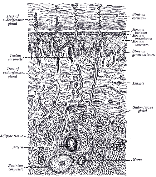
The skin is the largest organ in the body, covering roughly 1.8 square metres and accounting for about 15 percent of body weight. It is not a passive wrapper. It is a self-repairing barrier against water loss, mechanical damage, ultraviolet radiation, and microorganisms; a sensory sheet reporting touch, temperature, and injury; the main effector of thermoregulation; and an endocrine organ that begins vitamin D synthesis.
Anatomy: the structure
Three layers make up the skin and its support. The epidermis is avascular stratified squamous keratinised epithelium of ectodermal origin, nourished by diffusion from below. The dermis is mesodermal connective tissue carrying blood vessels, lymphatics, nerves, and the skin appendages. The hypodermis, or subcutaneous layer, is adipose and loose connective tissue that insulates, cushions, and anchors skin to underlying fascia while allowing it to slide.
The epidermis is arranged in strata that record the life cycle of a single cell type. The stratum basale is a single layer of mitotically active cells on the basement membrane. The stratum spinosum shows the spiny appearance created by desmosomal junctions in fixed tissue. The stratum granulosum contains keratohyalin granules and lamellar bodies that release the lipid mixture forming the true water barrier. The stratum lucidum is a clear layer present only in thick skin of palms and soles. The stratum corneum is the outermost layer of dead, flattened, keratin-filled corneocytes embedded in that lipid matrix, often described as a brick and mortar arrangement.
The dermis has two zones and holds most of the skin's functional structures. The superficial papillary dermis is loose connective tissue that interdigitates with the epidermis through dermal papillae, increasing adhesion and carrying the capillary loops that feed the epidermis. The deeper reticular dermis is dense irregular connective tissue with thick collagen bundles and elastic fibres that provide tensile strength and recoil. Within the dermis sit hair follicles with their arrector pili muscles and sebaceous glands, eccrine sweat glands present over almost the whole body, apocrine glands in the axilla and groin, and encapsulated sensory receptors.
Physiology: how it works
Barrier function works in several directions at once. The lipid-rich stratum corneum limits transepidermal water loss to a few hundred millilitres a day, which is why extensive burns cause such severe fluid deficits. Melanin produced by melanocytes and transferred to keratinocytes forms supranuclear caps that absorb ultraviolet radiation and protect DNA. The acid mantle, a surface pH of roughly 4 to 6 maintained by sweat and sebum, together with antimicrobial peptides and the resident microbiota, limits colonisation by pathogens.
Thermoregulation depends on the dermal circulation and the eccrine glands. Cutaneous blood flow can vary between roughly 1 and 100 millilitres per 100 grams of tissue per minute; vasodilation carries core heat to the surface for radiative loss, and vasoconstriction, aided by arteriovenous anastomoses in the fingers, toes, and ears, conserves it. Evaporation of sweat removes about 0.58 kilocalories per millilitre and is the only effective mechanism once ambient temperature exceeds skin temperature. Hypothalamic thermoreceptors integrate core and skin temperature and drive both responses.
Sensation is handled by receptors matched to different stimuli. Meissner corpuscles in the dermal papillae detect light touch and low-frequency vibration, Pacinian corpuscles deep in the dermis and hypodermis detect deep pressure and high-frequency vibration, Merkel cells at the basal layer signal sustained pressure and fine texture, and Ruffini endings detect skin stretch. Free nerve endings carry pain and temperature. Two-point discrimination varies from a couple of millimetres on the fingertip to several centimetres on the back, reflecting receptor density and cortical representation. Separately, ultraviolet B converts 7-dehydrocholesterol in the epidermis into cholecalciferol, the first step in vitamin D synthesis.
Biology: the living cells
Keratinocytes make up about 90 percent of epidermal cells, and their entire biology is a controlled programme of terminal differentiation. A basal cell divides, leaves the basement membrane, accumulates keratin filaments, produces keratohyalin and lamellar granules in the granular layer, then undergoes a specialised form of programmed cell death in which the nucleus and organelles are destroyed and the cell becomes a corneocyte, before finally being shed. The full journey takes about four weeks. Three non-keratinocyte populations share the epidermis: melanocytes in the basal layer, roughly one per ten basal keratinocytes, which package melanin into melanosomes and transfer them to surrounding cells, with skin colour reflecting melanosome size and distribution rather than melanocyte number; Langerhans cells, dendritic antigen-presenting cells in the spinous layer that sample the environment and migrate to lymph nodes; and Merkel cells in the basal layer, associated with sensory nerve terminals. In the dermis, fibroblasts produce collagen, mainly type I, and elastin, mast cells mediate immediate hypersensitivity, and macrophages provide surveillance and repair.
Histology: under the microscope
The distinction between thick and thin skin is the first thing to establish under the microscope, and the terms refer to epidermal thickness rather than overall skin thickness. Thick skin, found only on palms and soles, has a well-developed stratum lucidum, a very thick stratum corneum, prominent dermal papillae, abundant eccrine glands, and no hair follicles or sebaceous glands. Thin skin covers the rest of the body, lacks a stratum lucidum, has a much thinner corneum, and carries hair follicles with associated sebaceous glands. Sebaceous glands are recognisable as pale, foamy holocrine acini emptying into the follicle, since the entire cell disintegrates to release sebum. Eccrine sweat glands appear as coiled tubules in the deep dermis with a distinctive double-layered duct epithelium, and their myoepithelial cells help expel secretion. Beneath the epidermis, the papillary dermis stains lighter because its collagen is fine and loosely arranged, while the reticular dermis appears denser and more eosinophilic.
Clinical correlations
Burn severity is described by depth, and the anatomy predicts the outcome. Superficial burns involve only the epidermis and heal without scar. Superficial partial thickness burns reach the papillary dermis, blister, and heal from surviving appendages. Deep partial thickness burns reach the reticular dermis and heal slowly with scarring. Full thickness burns destroy the entire dermis including the appendages, so there is no source of re-epithelialisation and grafting is required. Extensive burns are dangerous mainly because the barrier is gone, producing massive fluid loss and a route for infection.
Skin cancer follows ultraviolet damage to keratinocyte and melanocyte DNA. Basal cell carcinoma arises from the basal layer, is the most common human cancer, and is locally invasive but rarely metastasises. Squamous cell carcinoma arises from keratinocytes of the spinous layer and carries a real though modest metastatic risk. Melanoma arises from melanocytes and, because those cells sit at the basal layer adjacent to dermal lymphatics and vessels, depth of invasion measured as Breslow thickness is the single strongest prognostic factor.
Wound healing proceeds through haemostasis, inflammation, proliferation, and remodelling, and remodelling continues for a year or more as type III collagen is replaced by type I. Scar tissue never regains full tensile strength, reaching roughly 80 percent of the original at best. Chronic inflammatory conditions map onto the layers as well: psoriasis is a disorder of accelerated keratinocyte turnover in which transit time falls from about four weeks to a few days, producing parakeratosis and a thickened plaque, while eczema reflects barrier dysfunction with intercellular oedema in the epidermis.
Common questions about the skin
What are the layers of the epidermis in order?
From deep to superficial: stratum basale, stratum spinosum, stratum granulosum, stratum lucidum in thick skin only, and stratum corneum. Each layer represents a stage in the maturation of a keratinocyte as it moves toward the surface and is eventually shed.
What is the difference between thick and thin skin?
The terms describe epidermal thickness. Thick skin occurs only on the palms and soles, has a stratum lucidum and a substantial stratum corneum, and contains no hair follicles or sebaceous glands. Thin skin covers everywhere else, lacks a stratum lucidum, and does carry hair follicles and sebaceous glands.
How does skin control body temperature?
Dermal blood vessels dilate to bring core heat to the surface for radiation and convection, or constrict to conserve it. Eccrine sweat glands add evaporative cooling, which becomes the only effective route once the surrounding air is warmer than the skin. The hypothalamus coordinates both responses.
Why do deep wounds scar while shallow ones do not?
Shallow injuries leave hair follicles and sweat glands intact, and epithelium regenerates from these appendages with normal architecture. Injuries that destroy the reticular dermis and its appendages remove that source, so the defect fills with fibroblast-produced collagen laid down in a disorganised pattern, which is scar.
Selected references
- Standring S, ed. Gray's Anatomy: The Anatomical Basis of Clinical Practice. 42nd ed. Elsevier; 2021.
- Ross MH, Pawlina W. Histology: A Text and Atlas. 8th ed. Wolters Kluwer; 2020.
- Hall JE, Hall ME. Guyton and Hall Textbook of Medical Physiology. 14th ed. Elsevier; 2021.
- Elias PM. Stratum corneum defensive functions: an integrated view. Journal of Investigative Dermatology. 2005;125(2):183-200.
- Bolognia JL, Schaffer JV, Cerroni L. Dermatology. 5th ed. Elsevier; 2024.
- Mescher AL. Junqueira's Basic Histology: Text and Atlas. 16th ed. McGraw Hill; 2021.
Medical disclaimer. This article is written for education and general understanding. It is not medical advice and cannot replace assessment by a qualified healthcare professional. If you have symptoms or health concerns, speak with a clinician. Read the full disclaimer.